

A symmetrical tetramer for S. aureus pyruvate carboxylase in complex with coenzyme A
- PMID: 19523900
- PMCID: PMC2731552
- DOI: 10.1016/j.str.2009.04.008
A symmetrical tetramer for S. aureus pyruvate carboxylase in complex with coenzyme A
Abstract
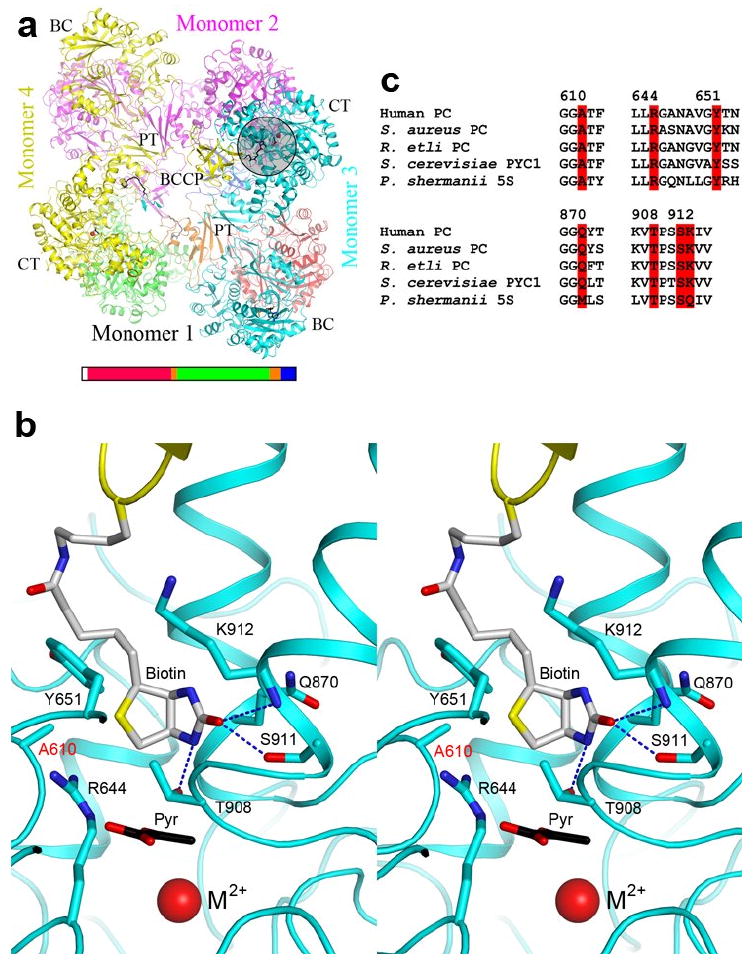
Pyruvate carboxylase (PC) is a conserved metabolic enzyme with important cellular functions. We report crystallographic and cryo-electron microscopy (EM) studies of Staphylococcus aureus PC (SaPC) in complex with acetyl-CoA, an allosteric activator, and mutagenesis, biochemical, and structural studies of the biotin binding site of its carboxyltransferase (CT) domain. The disease-causing A610T mutation abolishes catalytic activity by blocking biotin binding to the CT active site, and Thr908 might play a catalytic role in the CT reaction. The crystal structure of SaPC in complex with CoA reveals a symmetrical tetramer, with one CoA molecule bound to each monomer, and cryo-EM studies confirm the symmetrical nature of the tetramer. These observations are in sharp contrast to the highly asymmetrical tetramer of Rhizobium etli PC in complex with ethyl-CoA. Our structural information suggests that acetyl-CoA promotes a conformation for the dimer of the biotin carboxylase domain of PC that might be catalytically more competent.
Figures


Similar articles
-
Interaction between the biotin carboxyl carrier domain and the biotin carboxylase domain in pyruvate carboxylase from Rhizobium etli.Biochemistry. 2011 Nov 15;50(45):9708-23. doi: 10.1021/bi201277j. Epub 2011 Oct 18. Biochemistry. 2011. PMID: 21958016 Free PMC article.
-
Allosteric regulation of the biotin-dependent enzyme pyruvate carboxylase by acetyl-CoA.Biochem Soc Trans. 2012 Jun 1;40(3):567-72. doi: 10.1042/BST20120041. Biochem Soc Trans. 2012. PMID: 22616868 Review.
-
Characterizing the importance of the biotin carboxylase domain dimer for Staphylococcus aureus pyruvate carboxylase catalysis.Biochemistry. 2013 Jan 22;52(3):488-96. doi: 10.1021/bi301294d. Epub 2013 Jan 9. Biochemistry. 2013. PMID: 23286247 Free PMC article.
-
Crystal structures of human and Staphylococcus aureus pyruvate carboxylase and molecular insights into the carboxyltransfer reaction.Nat Struct Mol Biol. 2008 Mar;15(3):295-302. doi: 10.1038/nsmb.1393. Epub 2008 Feb 24. Nat Struct Mol Biol. 2008. PMID: 18297087
-
Regulation of the structure and activity of pyruvate carboxylase by acetyl CoA.Arch Biochem Biophys. 2012 Mar 15;519(2):118-30. doi: 10.1016/j.abb.2011.11.015. Epub 2011 Nov 19. Arch Biochem Biophys. 2012. PMID: 22120519 Free PMC article. Review.
Cited by
-
Structural insight into synergistic activation of human 3-methylcrotonyl-CoA carboxylase.Nat Struct Mol Biol. 2025 Jan;32(1):73-85. doi: 10.1038/s41594-024-01379-3. Epub 2024 Sep 2. Nat Struct Mol Biol. 2025. PMID: 39223421
-
Probing the allosteric activation of pyruvate carboxylase using 2',3'-O-(2,4,6-trinitrophenyl) adenosine 5'-triphosphate as a fluorescent mimic of the allosteric activator acetyl CoA.Arch Biochem Biophys. 2011 May 15;509(2):117-26. doi: 10.1016/j.abb.2011.03.006. Epub 2011 Mar 21. Arch Biochem Biophys. 2011. PMID: 21426897 Free PMC article.
-
Specificity and selectivity in post-translational biotin addition.Biochem Soc Trans. 2018 Dec 17;46(6):1577-1591. doi: 10.1042/BST20180425. Epub 2018 Oct 31. Biochem Soc Trans. 2018. PMID: 30381340 Free PMC article. Review.
-
Insights into the carboxyltransferase reaction of pyruvate carboxylase from the structures of bound product and intermediate analogs.Biochem Biophys Res Commun. 2013 Nov 15;441(2):377-82. doi: 10.1016/j.bbrc.2013.10.066. Epub 2013 Oct 22. Biochem Biophys Res Commun. 2013. PMID: 24157795 Free PMC article.
-
The three-dimensional structure of the biotin carboxylase-biotin carboxyl carrier protein complex of E. coli acetyl-CoA carboxylase.Structure. 2013 Apr 2;21(4):650-7. doi: 10.1016/j.str.2013.02.001. Epub 2013 Mar 14. Structure. 2013. PMID: 23499019 Free PMC article.
References
-
- Attwood PV. The structure and the mechanism of action of pyruvate carboxylase. Int J Biochem Cell Biol. 1995;27:231–249. - PubMed
-
- Attwood PV, Wallace JC. Chemical and catalytic mechanisms of carboxyl transfer reactions in biotin-dependent enzymes. Acc Chem Res. 2002;35:113–120. - PubMed
-
- Baxter WT, Leith A, Frank J. SPIRE: the SPIDER reconstruction engine. J Struct Biol. 2007;157:56–63. - PubMed
Publication types
MeSH terms
Substances
Grants and funding
LinkOut - more resources
Full Text Sources
Other Literature Sources
Molecular Biology Databases
