

Early keratocyte apoptosis after epithelial scrape injury in the human cornea
- PMID: 19523947
- PMCID: PMC2743797
- DOI: 10.1016/j.exer.2009.06.003
Early keratocyte apoptosis after epithelial scrape injury in the human cornea
Abstract
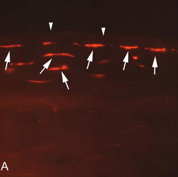
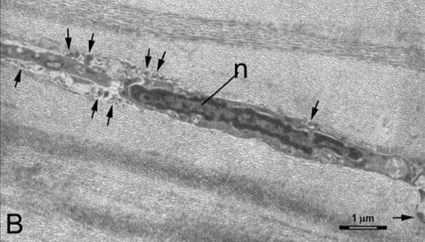
Animal studies in mice, rats, rabbits, pigs and hens demonstrated that anterior keratocytes undergo programmed cell death or apoptosis after corneal epithelial injury. Many other wound healing changes subsequently follow the keratocyte apoptosis response. This study evaluated early keratocyte apoptosis after corneal epithelial scrape injury in human eyes scheduled for enucleation for malignancy. Two eyes had corneal epithelial scrape 1 h prior to the enucleation and another eye served as a control and had no corneal scrape prior to enucleation. One additional eye was enucleated, washed with balanced salt solution, and then had the corneal epithelium scraped 1 h prior to processing for analysis. Apoptosis was identified by terminal deoxynucleotidyl transferase-mediated dUTP nick end labeling (TUNEL) assay and confirmed by transmission electron microscopy (TEM). Anterior keratocyte apoptosis was detected in the three corneas that had epithelial scrape injury, but not in the control unwounded cornea. This study confirmed that keratocyte apoptosis is also an early response to corneal epithelial injury in humans and showed that tears are not essential for keratocyte apoptosis to occur in response to epithelial injury.
Figures
References
-
- Arends MJ, Wyllie AH. Apoptosis: mechanisms and roles in pathology. Int Rev Exp Pathol. 1991;32:223–54. - PubMed
-
- Campos M, Szerenyi K, Lee M, McDonnell JM, McDonnell PJ. Keratocyte loss after corneal deepithelialization in primates and rabbits. Arch Ophthalmol. 1994;112:254–260. - PubMed
-
- Dohlman CH, Gasset AR, Rose J. The effect of the absence of corneal epithelium or endothelium on stromal keratocytes. Invest Ophthalmol Vis Sci. 1968;7:520–534. - PubMed
-
- Gao J, Gelber-Schwalb TA, Addeo JV, Stern ME. Apoptosis in the rabbit cornea after photorefractive keratectomy. Cornea. 1997;16:200–208. - PubMed
Publication types
MeSH terms
Grants and funding
LinkOut - more resources
Full Text Sources
Medical
