

Expression of the diabetes risk gene wolframin (WFS1) in the human retina
- PMID: 19523951
- PMCID: PMC2788494
- DOI: 10.1016/j.exer.2009.05.007
Expression of the diabetes risk gene wolframin (WFS1) in the human retina
Abstract
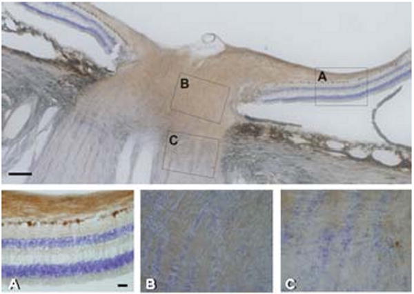
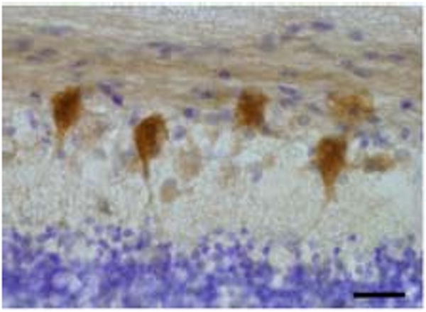
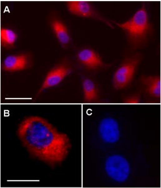
Wolfram syndrome 1 (WFS1, OMIM 222300), a rare genetic disorder characterized by optic nerve atrophy, deafness, diabetes insipidus and diabetes mellitus, is caused by mutations of WFS1, encoding WFS1/wolframin. Non-syndromic WFS1 variants are associated with the risk of diabetes mellitus due to altered function of wolframin in pancreatic islet cells, expanding the importance of wolframin. This study extends a previous report for the monkey retina, using immunohistochemistry to localize wolframin on cryostat and paraffin sections of human retina. In addition, the human retinal pigment epithelial (RPE) cell line termed ARPE-19 and retinas from both pigmented and albino mice were studied to assess wolframin localization. In the human retina, wolframin was expressed in retinal ganglion cells, optic axons and the proximal optic nerve. Wolframin expression in the human retinal pigment epithelium (RPE) was confirmed with intense cytoplasmic labeling in ARPE-19 cells. Strong labeling of the RPE was also found in the albino mouse retina. Cryostat sections of the mouse retina showed a more extended pattern of wolframin labeling, including the inner nuclear layer (INL) and photoreceptor inner segments, confirming the recent report of Kawano et al. [Kawano, J., Tanizawa, Y., Shinoda, K., 2008. Wolfram syndrome 1 (Wfs1) gene expression in the normal mouse visual system. J. Comp. Neurol. 510, 1-23]. Absence of these cells in the human specimens despite the use of human-specific antibodies to wolframin may be related to delayed fixation. Loss of wolframin function in RGCs and the unmyelinated portion of retinal axons could explain optic nerve atrophy in Wolfram Syndrome 1.
Figures

References
-
- Al-Till M, Jarrah NS, Ajlouni KM. Ophthalmologic findings in fifteen patients with Wolfram syndrome. Eur J Ophthalmol. 2002;12:84–88. - PubMed
-
- Awai M, Koga T, Inomata Y, Oyadomari S, Gotoh T, Mori M, Tanihara H. NMDA-induced retinal injury is mediated by an endoplasmic reticulum stress-related protein, CHOP/GADD153. J Neurochem. 2006;96:43–52. - PubMed
-
- Barrett TG, Bundey SE, Fielder AR, Good PA. Optic atrophy in Wolfram (DIDMOAD) syndrome. Eye. 1997;11:882–888. - PubMed
Publication types
MeSH terms
Substances
Grants and funding
LinkOut - more resources
Full Text Sources
Miscellaneous
