

Review
doi: 10.1016/j.tcb.2009.03.009.
Epub 2009 Jun 11.
Emerging roles for myosin II and cytoplasmic dynein in migrating neurons and growth cones
Affiliations
- PMID: 19524440
- PMCID: PMC2844727
- DOI: 10.1016/j.tcb.2009.03.009
Item in Clipboard
Review
Emerging roles for myosin II and cytoplasmic dynein in migrating neurons and growth cones
Trends Cell Biol.
2009 Jul.
Abstract
Motor proteins are involved in a wide range of cellular and subcellular movements. Recent studies have implicated two motor proteins in particular, myosin II and cytoplasmic dynein, in diverse aspects of cell migration. This review focuses on emerging roles for these proteins in the nervous system, with particular emphasis on migrating neurons and neuronal growth cones. The former cells exhibit unusual features of centrosome and nuclear movement, whereas growth cones offer an opportunity to evaluate motor protein function in a region of cytoplasm free of these organelles.
Figures
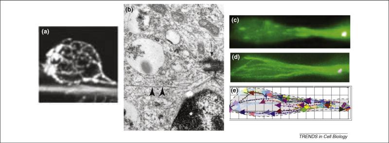
Substructure of migratory bipolar migrating neurons. (a) Anti-tubulin staining shows a cage of microtubules coursing around the nucleus of a cerebellar granule cell as it migrates along the process of an underlying radial glial cell [6]. (b) An electron micrograph of a neuronal precursor cell showing the centrosome (arrow) with radiating microtubules (arrowheads) located near the nucleus (indicated by asterisk) [5]. (c) Microtubule plus ends decorated with GFP–EB3 in live bipolar neuronal precursor cell located within embryonic rat brain slice [9]. (d) Superposition of movie frames during 2 minute imaging period and (e) tracings showing path and orientation of microtubules [9].Part(a) reproduced, with permission, from Ref. [6]; part (b) reproduced, with permission, from Ref. [5]; and part (c) reproduced, with permission, from Ref. [9].
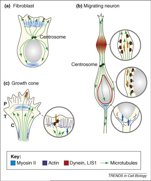
Diagrammatic representation of myosin II and cytoplasmic dynein roles in cell migration. Regions of (a) fibroblast, (b) migrating neuron and (c) growth cone in which myosin II and dynein are concentrated during migration are shown, along with actin filaments and microtubules. Microtubule plus ends are indicated by arrows. Actin filaments in lamellipodia and growth cone are primarily oriented with their plus ends toward the leading cell edge. In the expanded section, myosin II thick filaments are shown as light blue bars, dynein molecules in red. In fibroblasts and growth cones, myosin II concentrates in the transition zone (T) and produces a contractile force on growing filaments in the peripheral zone (P) extending from the leading cell edge. Myosin II also concentrates at the rear of the migrating fibroblast and at the growth cone neck. Cytoplasmic dynein and LIS1 (not shown) become concentrated within the leading lamellipodium and growth cone peripheral domain along with invading microtubules. In migrating neurons, myosin II acts behind the nucleus, which it seems to push forward. Cytoplasmic dynein acts from swellings that appear transiently within the migratory process and probably also from the nuclear surface. The centrosome is situated ahead of the nucleus in migrating fibroblast and neuron, but experiences force from dynein and LIS1 acting on microtubules well in advance. Arrows indicate direction of force production by dynein (orange) and myosin II (blue).
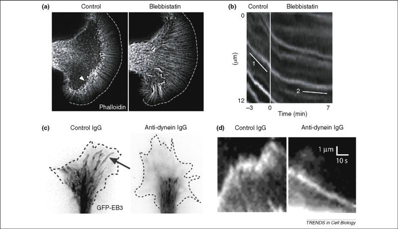
Effect of myosin II and dynein inhibition on growth-cone actin and microtubule behavior. (a) Slow-advancing Aplysia growth cone injected with Alexa594-phalloidin to image actin filaments. The myosin II inhibitor blebbistatin causes distortion of actin bundles and loss of actin arcs in the transition zone (indicated by arrowhead in control image). (b) Kymographs from live recording of the transition zone showing retrograde movement of phalloidin speckles (slope = line 1), which declines upon blebbistatin treatment (line 2). These data provide direct support for a permissive role for myosin II in retrograde actin flow. Parts (a) and (b) reproduced, with permission, from Ref. [34]. (c) Fast-advancing chicken dorsal root ganglia (DRG) growth cones plated on laminin and expressing GFP–EB3, which associates with the plus ends of growing microtubules. Images from video (30 frames/min) are overlaid and shown in negative contrast. Control microtubules (arrow) grow through the peripheral (P)-domain towards the leading growth-cone edge, behavior that is blocked by injected monoclonal antibody against dynein. (d) Kymographs show individual microtubule ends, revealed to be growing by the presence of GFP–EB3. The control microtubule advances and pauses as it continues to assemble as judged by the persistence of EB3 at the microtubule end. The microtubule from the dynein-inhibited growth cone moves in a retrograde direction. Persistence of EB3 at microtubule end again reveals that the microtubule continues to assemble. The rate of forward EB3 movement is that expected for microtubule assembly and is much slower than that for anterograde microtubule translocation. The rearward movement of microtubules in the dynein-inhibited cell provides direct evidence that dynein is responsible for restraining microtubules against rearward forces, such as retrograde actin flow. Parts (c) and (d) reproduced, with permission, from Ref. [17].
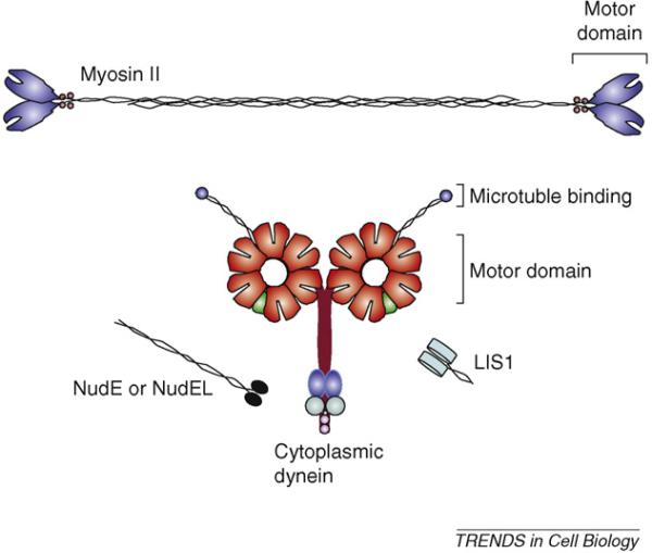
Structural organization of motor and regulatory proteins involved in neuronal migration and growth-cone motility. Color code: red, AAA ATPase modules; green, C-terminal regulatory domain; aqua, accessory cargo-binding intermediate chains; light purple, light chains; blue, light intermediate chains.
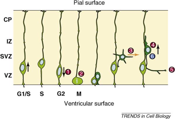
Stages in neurogenesis and migration in the developing neocortex. Abbreviations: CP, cortical plate; IZ, intermediate zone; SVZ, subventricular zone; VZ, ventricular zone.
References
Publication types
MeSH terms
Substances
Grants and funding
LinkOut - more resources
Full Text Sources
