. 2009 Jun 12;137(6):1018-31.
doi: 10.1016/j.cell.2009.03.051.

Retinoblastoma has properties of a cone precursor tumor and depends upon cone-specific MDM2 signaling

Affiliations

Retinoblastoma has properties of a cone precursor tumor and depends upon cone-specific MDM2 signaling

Xiaoliang L Xu et al. Cell. .

Abstract

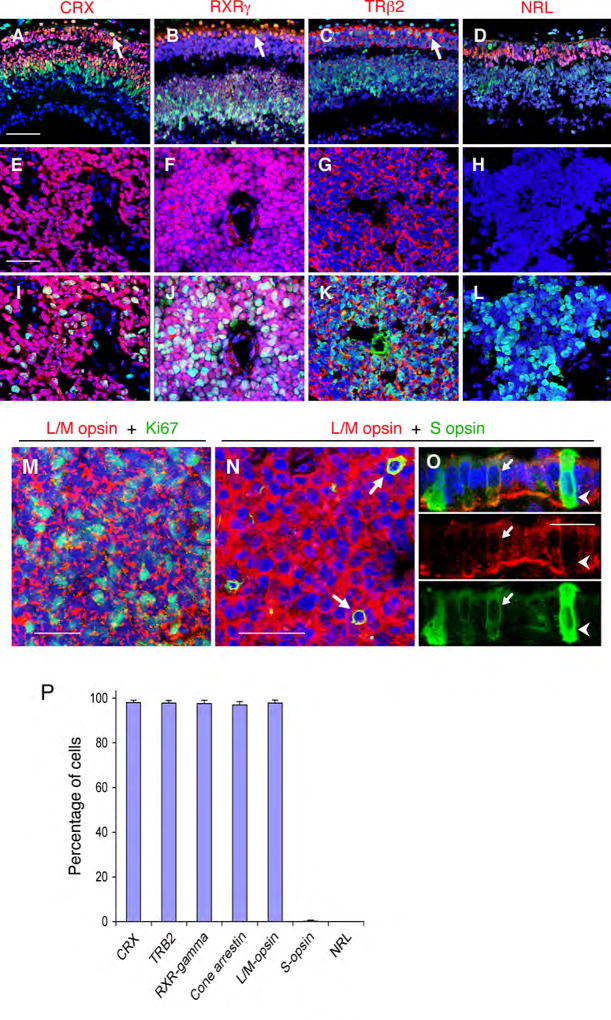
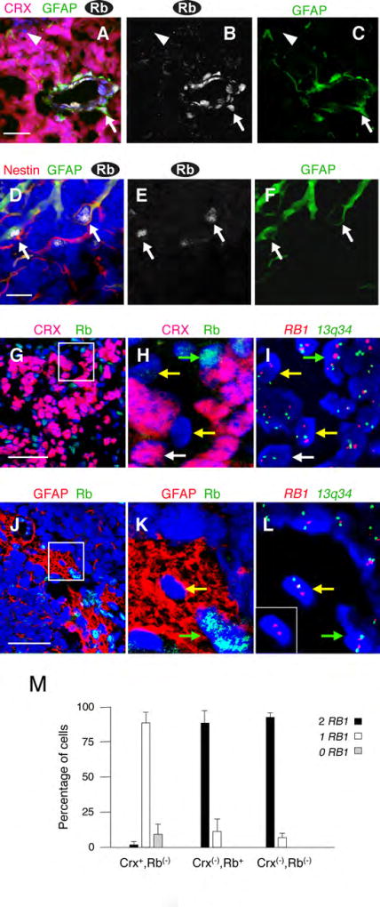
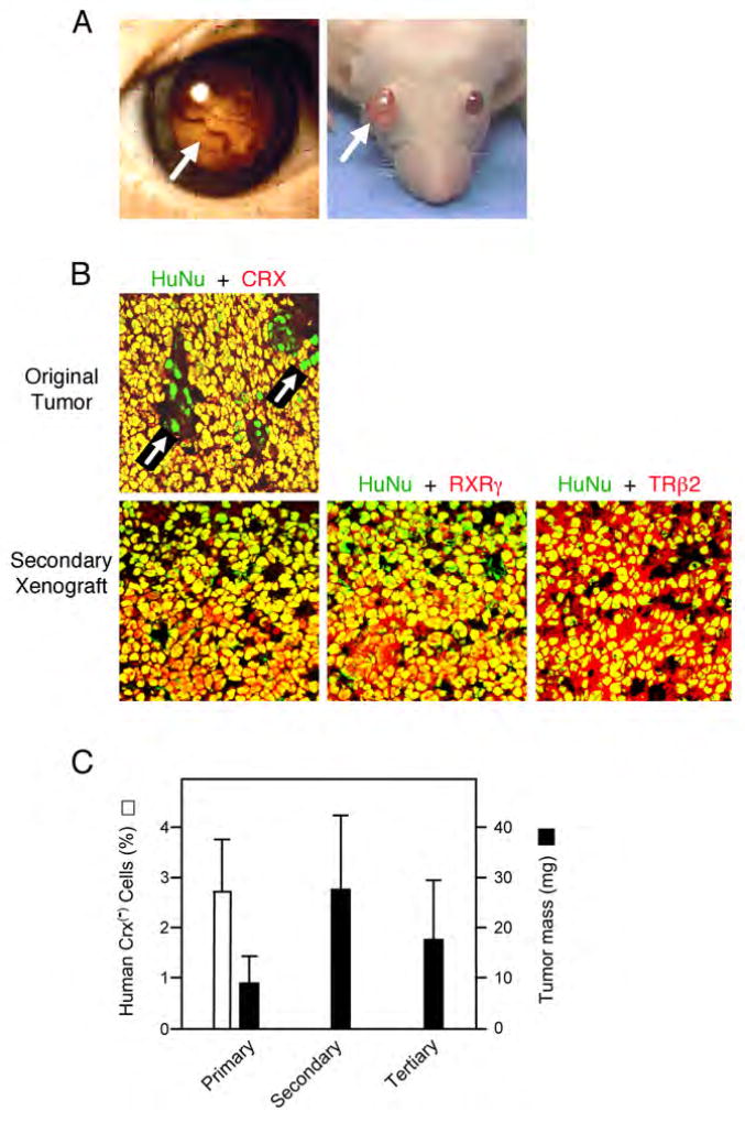
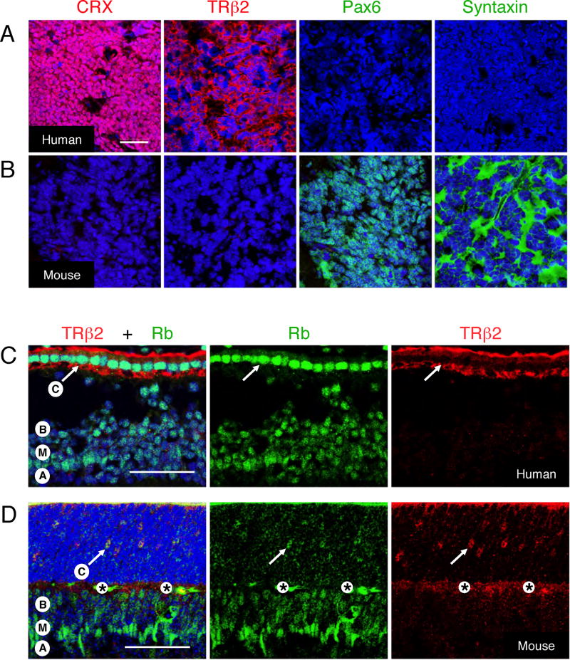
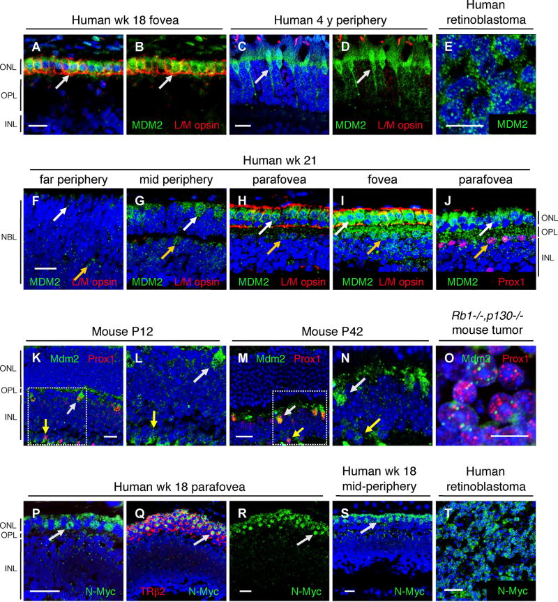
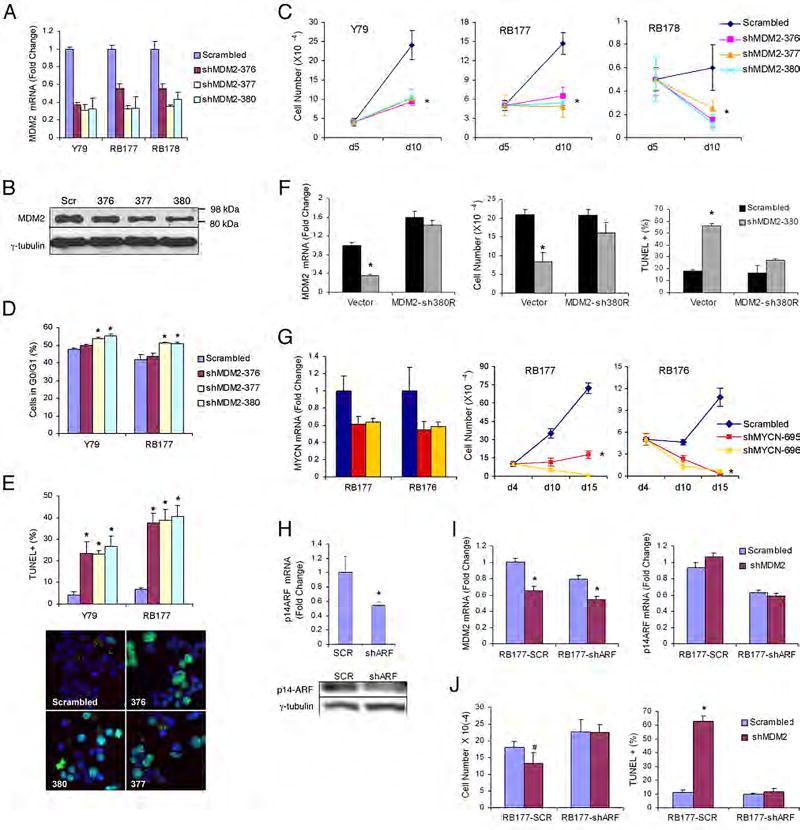
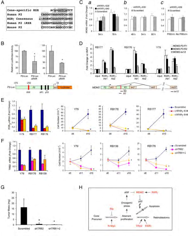
Retinoblastomas result from the inactivation of the RB1 gene and the loss of Rb protein, yet the cell type in which Rb suppresses retinoblastoma and the circuitry that underlies the need for Rb are undefined. Here, we show that retinoblastoma cells express markers of postmitotic cone precursors but not markers of other retinal cell types. We also demonstrate that human cone precursors prominently express MDM2 and N-Myc, that retinoblastoma cells require both of these proteins for proliferation and survival, and that MDM2 is needed to suppress ARF-induced apoptosis in cultured retinoblastoma cells. Interestingly, retinoblastoma cell MDM2 expression was regulated by the cone-specific RXRgamma transcription factor and a human-specific RXRgamma consensus binding site, and proliferation required RXRgamma, as well as the cone-specific thyroid hormone receptor-beta2. These findings provide support for a cone precursor origin of retinoblastoma and suggest that human cone-specific signaling circuitry sensitizes to the oncogenic effects of RB1 mutations.


Figures

Figure 1
Figure 1. Cone precursor markers in developing retina and retinoblastoma tumors
(A–D) Photoreceptor precursor markers (red) and Rb (green) in gestational wk 16 human retina. Note co-expression of Rb with cone markers CRX, RXRγ, and TRβ2 but not with the rod marker NRL (arrows, see also Figure S1). (E–L) Cone but not rod precursor markers (red) in retinoblastomas without (E–H) or with (I–L) Ki67 co-staining. The specificity of TRβ2 staining was confirmed by knockdown analyses (Figure S1). (M) Co-expression of L/M opsin (red) and Ki67 (green) in retinoblastoma. (N–O) Co-expression of L/M opsin and S opsin (arrows) in retinoblastoma (N) and in wk 18 perifoveal L/M cone precursors, but not in S cone precursors (O). Scale bars, 50 µm in A-N; 20 µm in O. (P) Mean percentage of cells expressing each marker in representative sections of 10 tumors (see Table S1). Error bars indicate standard deviation.
Figure 2
Figure 2. Retinoblastoma cells express cone but not glial markers
(A–F) Rb expression in retinoblastoma glia. (A–C) Rb (white) in endothelial cells and GFAP+ perivascular glia (green, arrow), but not in CRX+ tumor cells (red). Rare cells lack Rb, CRX, and GFAP (arrowhead). (D–F) Rb (white) in non-perivascular cells that co-express GFAP (green) and Nestin (red, arrows). (G–M) RB1 FISH of CRX,Rb and GFAP+,Rbcells. Sections were co-stained for Rb and either CRX (G–H) or GFAP (J–K), and then probed by FISH (I, L), and nuclei with two control 13q34 signals (green) examined for RB1 (red). Boxed regions in G and J are magnified in H-I and K-L, and the inset in L shows the RB1 FISH for the cell in the center of the image. Arrows in G-L indicate CRX+Rb tumor cells (white), non-neoplastic Rb+ cells (green), and CRX,Rb or GFAP+,Rb cells (yellow). (M) The percentage of cells with the indicated CRX and Rb staining that have 2, 1, or 0 RB1 signals. Error bars indicate standard deviation for six sections from two tumors. Scale bars: 50 µm in A, G, J; 20 µm in D.
Figure 3
Figure 3. Cone precursor-like cells propagate retinoblastoma
(A) Human retinoblastoma and mouse subretinal xenografts (arrows). (B) Original retinoblastoma (top) and secondary xenograft (bottom) co-stained with human nuclear antigen (HuNu, green) and either CRX, RXRγ, or TRβ2 (red). Most cells in the original tumor co-stained with HuNu and CRX (yellow). Occasional green nuclei (arrows) represent HuNu+,CRX cells. Green HuNu+ human cells lacking CRX, RXRγ, or TRβ2 were not detected in xenografts. (C) Percentage of human cells that lack CRX in samples used in primary, secondary and tertiary xenografts (□) and average mass of primary, secondary, and tertiary xenografts (■). Error bars indicate standard deviation.
Figure 4
Figure 4. Species-specific retinal tumor phenotypes and Rb expression patterns
(A–B) Human retinoblastoma (A) and a Rb1−/−,p130−/− retinal tumor (B) stained for cone markers CRX and TRβ2 (red), or for amacrine and horizontal cell markers syntaxin and Pax6 (green), and with DAPI (blue), using antibodies that stain the appropriate mouse and human retinal cells (Figure S8). (C–D) Prominent Rb (green) in TRβ2+ cone precursors (arrows) in human wk 18 fovea (C), but not in mouse P12 central retina (D). Labels indicate positions of cone (C), bipolar (B), Müller (M), and amacrine (A) cells, and an antibody-independent signal (*). Note that the P12 cone precursors in (D) are appropriately positioned in the outer nuclear layer (Rich et al., 1997; Ng et al., 2009). Scale bars, 50 µm.
Figure 5
Figure 5. MDM2 and N-Myc expression in human cone precursors
(A–J) MDM2 (green) in human gestational wk 18 (A, B) or wk 21 (F–J) retina, an uninvolved retina from a 4-year old retinoblastoma patient (C–D), or a retinoblastoma tumor (E); and co-stained for L/M opsin (A–D and F–I, red) or Prox1 (J, red). The same section was imaged with fixed parameters in F–I. White and yellow arrows indicate MDM2+ cones and horizontal cells, respectively. (K–O) Mdm2 (green) and Prox1 (red) in mouse P12 (K–L) or P42 (M–N) retina, or in a Rb1−/−,p130−/− mouse retinal tumor (O). Boxed regions in K and M are shown at higher magnification in L and N. White and yellow arrows indicate Mdm2+ horizontal and amacrine cells, respectively. (P–T) N-Myc (green) in wk 18 parafovea (P–R) or mid-periphery (S), or in human retinoblastoma (T), with TRβ2 co-staining (Q, red). Arrows indicate N-Myc+ cones. Scale bars, 20 µm for all panels except 10 µm in E and O.
Figure 6
Figure 6. Roles for MDM2 and N-Myc in retinoblastoma cells
(A) qRT-PCR of MDM2 RNA, 5 days after infection of Y79, RB177, or RB178 cells with MDM2 shRNAs or a scrambled control. (B) Immunoblot analysis of MDM2 and γ-tubulin (as a control) in Y79 cells on day 5 after infection. (C) Growth of Y79, RB177, and RB178 cells after puromycin selection, plating on day 5, and counting on days 5 and 10 after infection. (D) The proportion of G0/G1 cells in Y79 and RB177 cultures, 9 days after infection. (E) The percentage of TUNEL+ Y79 and RB177 cells, 8 days after infection (top) and representative TUNEL staining (bottom). (F) RB177 cells transduced with sh380 resistant MDM2 cDNA and re-infected with pLKO-shMDM2-380 or a scrambled shRNA control. Left, qRT-PCR analysis of MDM2 RNA, 4 days after re-infection. Middle, cell numbers after plating on day 4 and counting on day 10 after re-infection. Right, percentage of TUNEL+ cells 11 days after re-infection. (G) qRT-PCR analysis of MYCN RNA on day 5, and cell growth after plating on day 4 and counting on days 4, 10, and 15 after transduction of RB177 or RB176 with MYCN shRNAs. (H) RB177 cells transduced with pLKO-shARF-331 or a scrambled control, and analyzed for CDKN2AARF RNA (top) and ARF protein (bottom). (I) qRT-PCR analysis of MDM2 RNA and CDKN2AARF RNA, 4 days after re-infection of RB177-shARF and control cells with pLKO-shMDM2-377 or control shRNA. (J) Cell numbers and TUNEL staining 5 days after re-infection with MDM2 or control shRNA vectors. Error bars indicate standard deviation, and (*) or (#) indicate P < 0.01 or P = 0.016, respectively.
Figure 7
Figure 7. Roles for cone-specific transcription factors in retinoblastoma cellMDM2expression, proliferation, and survival
(A) A human MDM2 P2 promoter element (second row) with identity to a cone-specific RXR element (Danko et al., 2007) at each of six invariant positions (top row, underlined), and identity to the consensus RXRγ homodimer binding site at 14 of 15 positions (third row, shaded, from Table 1 of Dowhan et al., 1994), but differing from murine sequences (fifth row, aligned as in Figure S13). Human-to-mouse substitutions in P2-Luc-ΔRXR (fourth row) are underlined. (B) Top, luciferase activities after transfection of P2-Luc and P2-Luc-ΔRXR in two experiments. Bottom, P2-Luc structure. (C) qRT-PCR analysis of MDM2 RNA in RB177 cells at 54 and 78 h after transduction with RXRγ shRNAs (a); at 48, 54, and 60 h after transduction (b); and at 30 h after transduction in the presence or absence of 60 nM pifithrin-α. p-nitro, cyclic (EMD Biosciences) for the last 6h (c). (D) Top, ChIP analysis of RB177, RB176, and Y79 assayed by qRT-PCR directly (input) or after immunoprecipitation with two RXRγ antibodies. Values are the ratio of the MDM2 PCR products R1, R2, or ex12 to an HNF4α product. Bottom, the human MDM2 locus, showing positions of the RXR site and PCR products. (E) qRT-PCR analysis of RXRγ RNA on day 4, and cell growth after plating on day 4 after transduction of Y79, RB176, or RB177 with RXRγ shRNAs. (F) qRT-PCR analysis of TRβ2 RNA at day 5, and cell growth after plating on day 5 and counting on days 6, 11, and 16 after transduction of Y79, RB176, or RB139 cells with the indicated TRβ shRNAs. (G) Mean tumor mass 50 days after engrafting Y79 cells transduced with shTRβ2, shTRβ1+2, or scrambled shRNAs. (H) Potential role of cone precursor signaling proteins in retinoblastoma tumorigenesis. Proteins in Red are highly expressed during human cone precursor maturation. Error bars indicate standard deviation, and asterisks indicate P < 0.05.

Comment in

References

    1. Abramson DH, Gombos DS. The topography of bilateral retinoblastoma lesions. Retina. 1996;16:232–239. - PubMed
    1. Ajioka I, Martins RA, Bayazitov IT, Donovan S, Johnson DA, Frase S, Cicero SA, Boyd K, Zakharenko SS, Dyer MA. Differentiated horizontal interneurons clonally expand to form metastatic retinoblastoma in mice. Cell. 2007;131:378–390. - PMC - PubMed
    1. Bibb LC, Holt JK, Tarttelin EE, Hodges MD, Gregory-Evans K, Rutherford A, Lucas RJ, Sowden JC, Gregory-Evans CY. Temporal and spatial expression patterns of the CRX transcription factor and its downstream targets. Critical differences during human and mouse eye development. Hum Mol Genet. 2001;10:1571–1579. - PubMed
    1. Bogenmann E, Lochrie MA, Simon MI. Cone cell-specific genes expressed in retinoblastoma. Science. 1988;240:76–78. - PubMed
    1. Chen D, Livne-bar I, Vanderluit JL, Slack RS, Agochiya M, Bremner R. Cell-specific effects of RB or RB/p107 loss on retinal development implicate an intrinsically death-resistant cell-of-origin in retinoblastoma. Cancer Cell. 2004;5:539–551. - PubMed

Publication types

MeSH terms

Substances