

Review
doi: 10.1016/j.molcel.2009.05.016.
RVB1/RVB2: running rings around molecular biology
Affiliations
- PMID: 19524533
- PMCID: PMC2733251
- DOI: 10.1016/j.molcel.2009.05.016
Item in Clipboard
Review
RVB1/RVB2: running rings around molecular biology
Mol Cell.
.
Abstract
RVB1/RVB2 (also known as Pontin/Reptin, TIP49/TIP48, RuvbL1/RuvbL2, ECP54/ECP51, INO80H/INO80J, TIH1/TIH2, and TIP49A/TIP49B) are two highly conserved members of the AAA+ family that are present in different protein and nucleoprotein complexes. Recent studies implicate the RVB-containing complexes in many cellular processes such as transcription, DNA damage response, snoRNP assembly, cellular transformation, and cancer metastasis. In this review, we discuss recent advances in our understanding of RVB-containing complexes and their role in these pathways.
Figures
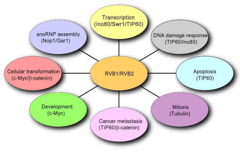
RVB1 and RVB2 are involved in multiple cellular pathways. Schematic showing involvement of RVBs in transcription, DNA damage response, small nucleolar ribonucleotide protein (snoRNPs) assembly, cellular transformation, cancer metastasis, apoptosis, mitosis and development. RVBs act in (i) transcription by regulating function of Ino80, Swr1 and TIP60 complexes, (ii) DNA damage response through TIP60 and Ino80, (iii) snoRNP assembly by affecting maturation of snoRNAs and localization of Nop1 and Gar1, (iv) cellular transformation by c-Myc and β-catenin function, (v) cancer metastasis by regulating expression of KAI1 through TIP60 and β-catenin, (vi) apoptosis through TIP60, (vii) mitosis by regulating assembly of microtubules and (viii) development through c-Myc pathway.
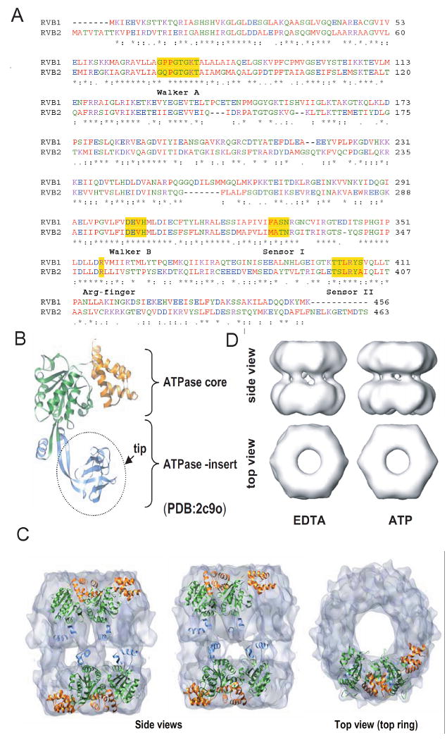
Sequence and structural details of RVBs. (A) Protein sequence alignment of human RVB1 and RVB2 showing regions of similarity where Walker A, Walker B, sensor 1, sensor 2 and arginine finger are highlighted. (B) Structure of monomeric RVB1. ATPase core domain consisting of domain I (1-120 aa + 296-365 aa) and domain III (368-456 aa) and ATPase insert domain II (121-295 aa) are shown. (C) Electron microscopic (EM) image of dodecameric RVB1 and RVB2. RVB1 and RVB2 are arranged as a double hexamer and interact through their ATPase insert domain. (D) Change in conformation after binding of ATP. Different views of the EM images of RVB1 and RVB2 in presence of ATP or EDTA.
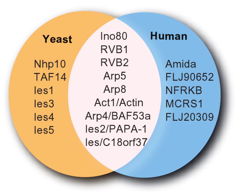
Composition of human and yeast Ino80 complexes. Subunits that are common and different between human and yeast Ino80 complex are shown.
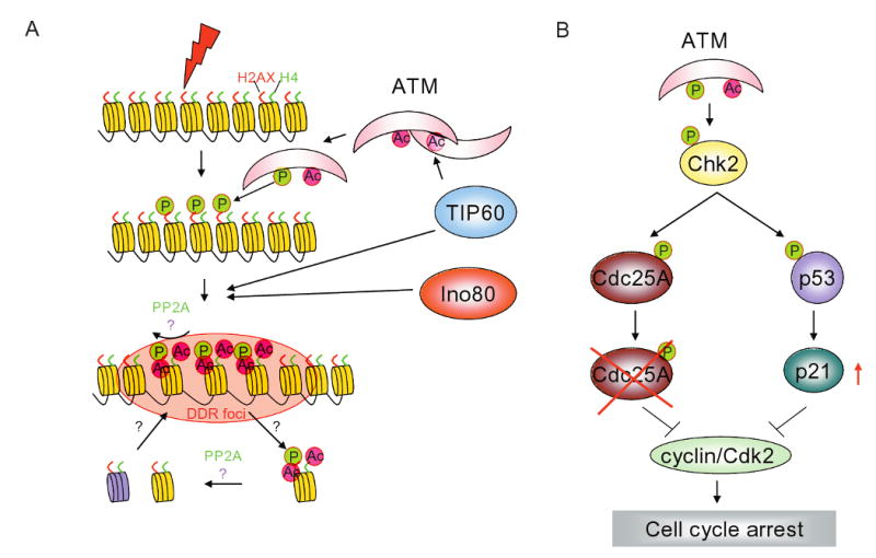
RVBs are involved at multiple steps in the DNA damage response pathways. (A) Schematic showing various steps of DNA damage response (DDR). Step 1) sensing and activation of DDR by sensors involves acetylation of ATM by TIP60 complex, 2) amplification of DDR signal by phosphorylation of H2AX by ATM and acetylation of histone H2AX and H4 by TIP60 complex, 3) recruitment of repair proteins at the site of damage is facilitated by chromatin de-condensation by action of TIP60 and Ino80 complexes. Relaxation of chromatin at the site of DNA damage enables binding of DNA damage repair protein and 4) signaling to activate cell cycle checkpoints. Activation of ATM results in phosphorylation of Chk2 that activates a fast and slow checkpoint signaling by either degrading Cdc25A or increasing levels of p21, respectively.
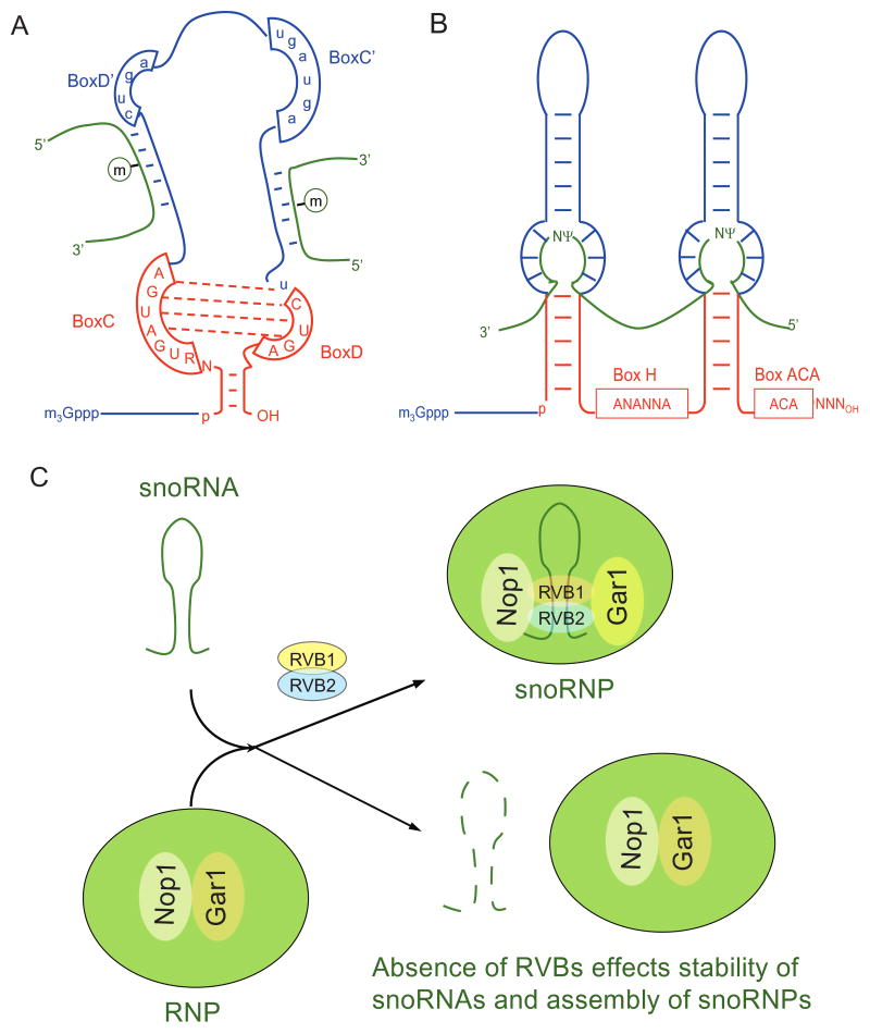
RVBs are required for small nucleoprotein (snoRNP) assembly. (A) and (B) Schematics of box C/D and H/ACA small nucleolar ribonuleotide (snoRNA) family. (C) RVB bind to the stem of snoRNA and are required maturation of snoRNAs. Loss of RVB affects proper localization of two core snoRNP proteins, Nop1 and Gar1 that associate with snoRNAs of box C/D family and H/ACA family, respectively.
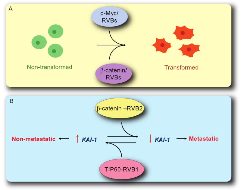
RVBs are involved in cellular transformation and cancer metastasis. (A) Cellular transformation by c-Myc and β-catenin requires functional RVBs. Expression of Walker B mutant of RVB1 acts as a dominant negative form of RVB1 and inhibits cellular transformation. (B) RVB1-TIP60 and RVB2-β-catenin complexes function antagonistically. RVB1-TIP60 complex act as an activator of KAI1 expression whereas RVB2-β-catenin complex act as a repressor. Inhibition of expression of KAI1 transforms cells from non-metaststic to metastatic state.
References
-
- Baek SH, Ohgi KA, Rose DW, Koo EH, Glass CK, Rosenfeld MG. Exchange of N-CoR corepressor and Tip60 coactivator complexes links gene expression by NF-kappaB and beta-amyloid precursor protein. Cell. 2002;110:55–67. - PubMed
-
- Bakkenist CJ, Kastan MB. DNA damage activates ATM through intermolecular autophosphorylation and dimer dissociation. Nature. 2003;421:499–506. - PubMed
Publication types
MeSH terms
Substances
Grants and funding
LinkOut - more resources
Full Text Sources
Other Literature Sources
Molecular Biology Databases
Miscellaneous
