. 2009 Jun 12;34(5):569-79.
doi: 10.1016/j.molcel.2009.04.028.

An unfolded CH1 domain controls the assembly and secretion of IgG antibodies

Affiliations

An unfolded CH1 domain controls the assembly and secretion of IgG antibodies

Matthias J Feige et al. Mol Cell. .

Abstract

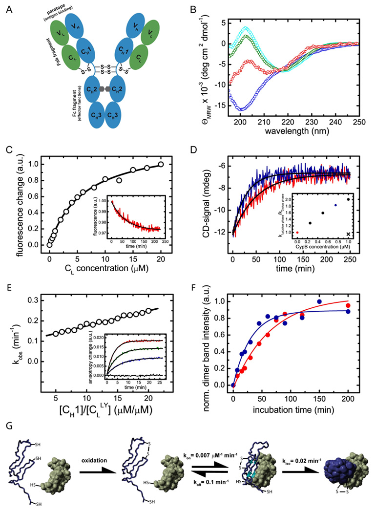
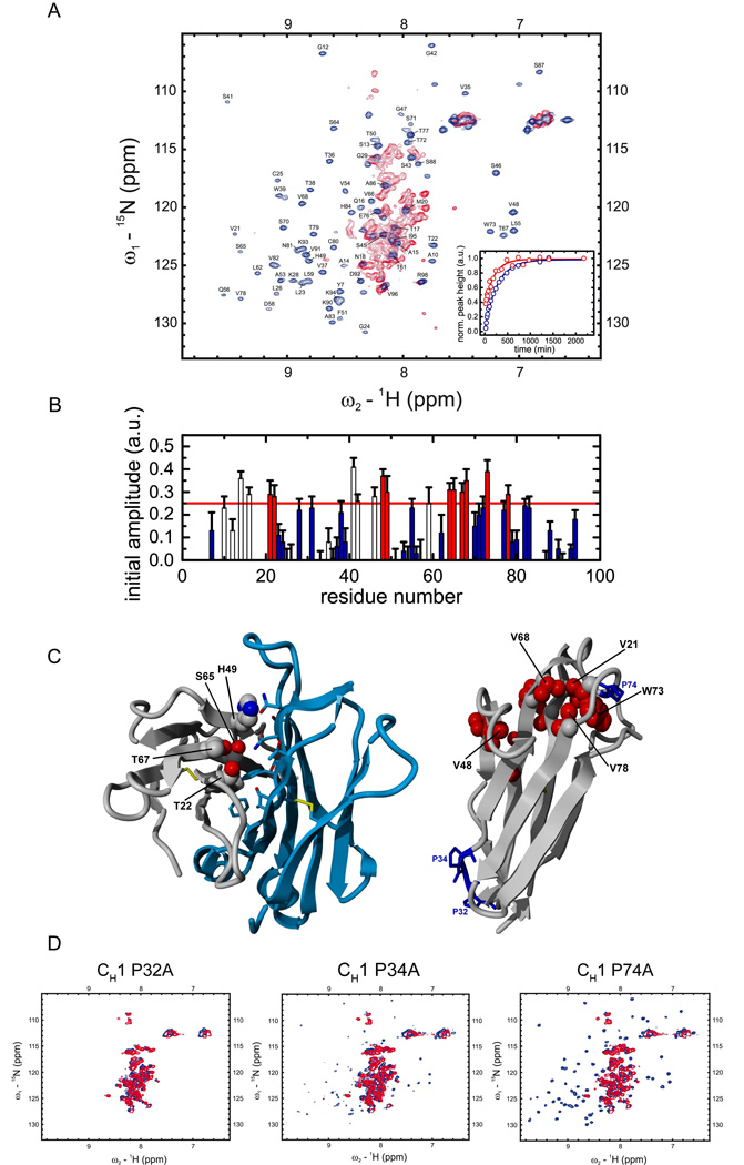
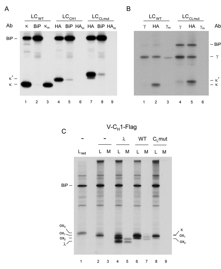
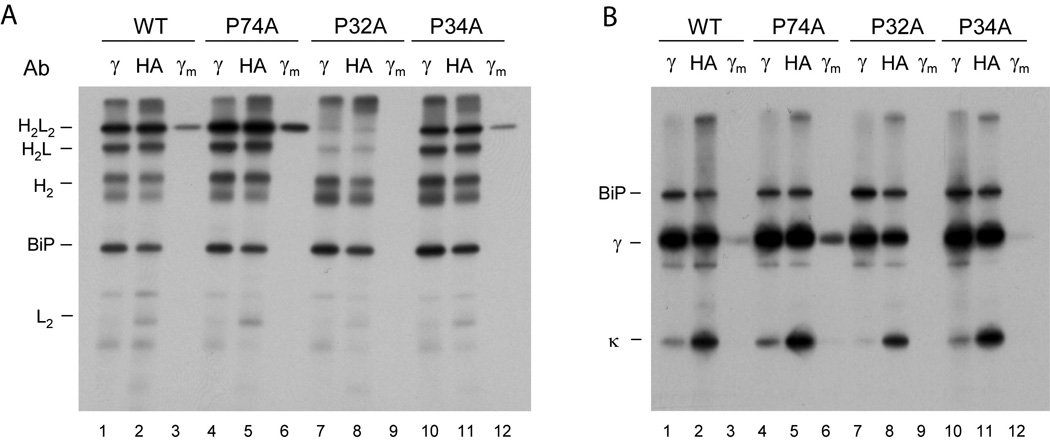
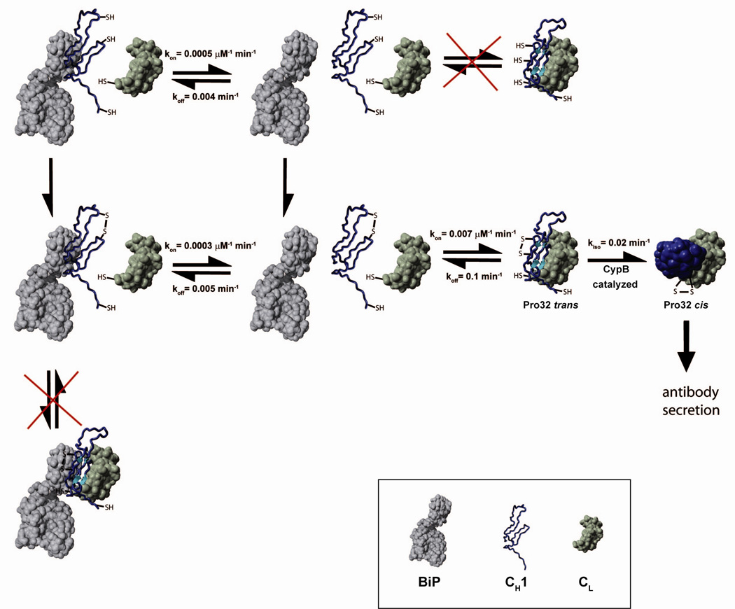
A prerequisite for antibody secretion and function is their assembly into a defined quaternary structure, composed of two heavy and two light chains for IgG. Unassembled heavy chains are actively retained in the endoplasmic reticulum (ER). Here, we show that the C(H)1 domain of the heavy chain is intrinsically disordered in vitro, which sets it apart from other antibody domains. It folds only upon interaction with the light-chain C(L) domain. Structure formation proceeds via a trapped intermediate and can be accelerated by the ER-specific peptidyl-prolyl isomerase cyclophilin B. The molecular chaperone BiP recognizes incompletely folded states of the C(H)1 domain and competes for binding to the C(L) domain. In vivo experiments demonstrate that requirements identified for folding the C(H)1 domain in vitro, including association with a folded C(L) domain and isomerization of a conserved proline residue, are essential for antibody assembly and secretion in the cell.


Conflict of interest statement

The authors declare no conflict of interest.

Figures

Figure 1
Figure 1. Structural characteristics of the CH1 domain and its assembly mechanism with the CL domain
(A) shows a schematic representation of the IgG1 molecule. The IgG molecule is composed of two heavy chains (blue) and two light chains (green). The heavy chain consists of the VH, CH1, CH2 and CH3 domain and the light chain of the VL and the CL domain (listing from N- to C-terminus of each chain). The CH2 domains of the heavy chains are glycosylated with complex biantennary oligosaccharides (depicted in grey). Each domain possesses an internal disulfide bridge (omitted for clarity) and additional disulfide bonds link the two heavy chains in the flexible hinge region. A single disulfide bond covalently connects CH1 with the CL domain. The two identical antigen binding sites (paratopes) are made up by the two variable domains VH and VL. The overall IgG molecule can be divided into two Fab fragments (composed of VH, CH1, VL and CL) and one Fc fragment (composed of two CH2 and two CH3 domains). (B) The isolated CL domain (cyan) displays a typical all-β far-UV CD spectrum whereas the isolated CH1 domain (blue) shows a random coil spectrum. To assess if CL induces structural changes in CH1, the spectrum of co-incubated CL and CH1 was recorded (green). From this spectrum and the far-UV CD spectrum of the isolated CL domain, the spectrum of the CH1 domain in the presence of CL was calculated (red) which shows the characteristics of β-sheet secondary structure. (C) The affinity between CH1 and CL was determined by the change in the intrinsic fluorescence upon CL induced folding of the CH1 domain, recorded before and after a 4 h equilibration step. A one-site binding model was used to fit the data. The inset shows a representative single exponential trace observed after the addition of 1 µM CL to 2 µM CH1. A single exponential reaction with a very similar rate was observed by far-UV CD spectroscopy (D, red trace). The folding reaction could be accelerated by the PPIase CypB (red trace: 10 µM CL and 10 µM CH1 alone, blue trace: in the presence of 0.75 µM CypB). The inset shows the dependence of the slow reaction on CypB concentration. The observed rate in the absence of CypB is denoted as k0, in the presence of CypB as kcat. If 1µM CypB was inhibited by 2 µM cyclosporine A, no acceleration was observed (black cross). (E) Association of the CH1 domain with a lucifer yellow labeled CL domain was followed by the change in the lucifer yellow anisotropy signal. The observed rate constants were fitted with a linear function to yield the kon value and the koff value of 0.007 ±0.0002 µM−1 min−1 respectively 0.1 ±0.01 min−1. The inset shows individual single exponential traces after the addition of 0 µM (black), 5 µM (blue), 10 µM (green) and 20 µM (red) CH1 to 1 µM labeled CL. (F) To assess the formation of the CL/CH1 interchain disulfide bridge, non-reducing SDS-PAGE experiments were carried out and the dimer band intensity was quantified. 25 µM of each domain were used. In the absence of CypB (red), a time constant of τ = 63 ±7 min was observed for the formation of covalent CL/CH1 dimers. In the presence of 5 µM CypB, a time constant of τ = 31 ±5 min was obtained. In (G), the overall CH1/CL assembly mechanism is shown. Only after formation of the internal disulfide bridge in the CH1 domain (blue), the fast formation of a dimeric intermediate with the CL domain (green) is observed. Subsequently, prolyl isomerization limits complete folding and formation of the interchain disulfide bridge. All measurements were carried out at 25°C in PBS.
Figure 2
Figure 2. NMR spectroscopic characterization of CL induced CH1 folding
(A) 15N-1H HSQC spectra of the isolated CH1 domain (red) and the assigned CH1 domain in complex with the CL domain (blue) are shown. In order to characterize the folding pathway of the intrinsically disordered CH1 domain, time dependent HSQC intensities upon addition of unlabeled CL to 15N labeled CH1 were measured for each assigned residue at the native chemical shift position and fitted by a single exponential function. Two representative traces for Val68 (red) and Lys90 (blue) are shown in the inset. (B) Initial amplitudes for each assigned CH1 residue were derived from the fitted exponential functions. Residues with an initial HSQC amplitude below a threshold of 25% native intensity are colored in blue and residues above the threshold in red (open bars: residues in loop regions / filled bars: residues in structured regions). Errors indicate standard deviations from single exponential fits. In (C) CH1 residues with intensities above the threshold in the intermediate are mapped on the crystal structure of the CH1/CL dimer (pdb code 1Q9K). The dimerization interface between CH1 (grey) and CL (blue) is shown on the left with only residues of CH1 indicated that are involved in this interaction and above the HSQC amplitude threshold. On the right, internal CH1 residues above the 25% threshold are shown in red, the three cis proline residues in CH1 are depicted and labeled in blue. (D) The HSQC spectra of the CH1 Pro32Ala, Pro34Ala and Pro74Ala mutants show, that only the Pro32Ala mutant is not able to fold in the presence of CL anymore (blue spectrum). For the other two mutants, Pro34Ala and Pro74Ala, well dispersed spectra and hence folding are observed in the presence of CL (blue spectra). In the absence of CL (red spectra), all three mutants show typical HSQC spectra of unfolded proteins. All measurements were carried out in PBS at 25°C except for the folding kinetics which where recorded at 12.5°C.
Figure 3
Figure 3. Characterization of the interaction between BiP and CH1 in vitro
(A) The affinity between BiP and oxidized CH1 (filled circles, straight line) as well as reduced CH1 (open circles, dashed line) was determined by analytical HPLC experiments. The data were fitted to a one-site binding model to determine the Kd. (B) The association kinetics between 1 µM BiP and varying concentrations of oxidized CH1 (filled circles) and reduced CH1 (open circles) were measured to determine the rate constants of the reaction. For oxidized CH1, kon = 0.00026 ±0.00002 µM−1 min−1 and koff = 0.0050 ±0.0002 min−1 were obtained. For the reduced CH1 domain, the corresponding values were kon = 0.00041 ±0.00003 µM−1 min−1 and koff = 0.0047 ±0.0003 min−1. The left inset shows single HPLC runs of 8 µM oxidized CH1 and 1 µM BiP after 10 min (blue) and 200 min (red) co-incubation. The right inset shows the overall observed single exponential association kinetics between 1 µM BiP and 8 µM oxidized CH1.
Figure 4
Figure 4. The folding status of an antibody domain controls IgG secretion in vivo
(A) COS-1 cells were co-transfected with vectors encoding BiP and either a wild type light chain (LCwt), a light chain containing the CH1 domain instead of the CL domain (LCCH1), or a light chain containing an unfolded CL domain (LCCLmut). Cells were metabolically labeled for 3 h and both cell lysates (no subscript) and culture supernatants (subscript m) were immunoprecipitated with the indicated antisera (Ab). Precipitated proteins were separated by SDS-PAGE under reducing conditions and visualized by autoradiography. (B) COS-1 cells were co-transfected as in (A) except that a vector encoding a chimeric humanized mouse heavy chain was also included. Cells were labeled and analyzed as in (A). (C) COS-1 cells were co-transfected with vectors encoding BiP, a Flag-tagged truncated heavy chain consisting of only the VH and CH1 domains (Lee et al., 1999), and with the indicated light chain constructs (i.e., λ, wild type κ, or CL mutant κ). Cells were metabolically labeled and both cell lysates (L) and culture supernatants (M) were immunoprecipitated with the anti-Flag antibody. Precipitated proteins were separated by SDS-PAGE under non-reducing condition (except the first lane, which included 2-ME in the sample buffer and is indicated as red) and visualized by autoradiography. Mobilities of completely reduced (ox0), partially oxidized (ox1), and fully oxidized (ox2) forms of truncated heavy chain, as well as those of λ and κ light chains are indicated. The tagged forms of the κ light chain constructs co-migrate with the ox1 form of the truncated heavy chain.
Figure 5
Figure 5. The isomerization of a single proline residue controls the assembly and secretion of heavy chains in vivo
COS-1 cells were co-transfected with vectors encoding wild type MAK33 heavy chain (WT) or one of the three Pro to Ala mutants (P74A, P32A, or P34A) together with wild type light chain and BiP. Cells were metabolically labeled and both cell lysates (no subscript) and culture supernatants (subscript m) were immunoprecipitated with the indicated antisera (Ab). Precipitated proteins were separated by SDS-PAGE under non-reducing conditions and visualized by autoradiography. (B) The cell lysates and culture supernatants from (A) were divided in half, immunoprecipitated with the indicated antibodies, analyzed by SDS-PAGE under reducing conditions, and visualized with autoradiography. These data demonstrate that the failure of the P32A mutant to induce assembly and secretion is not because it is expressed poorly, as the signal for this mutant heavy chain is very similar to that of the wild type heavy chain.
Figure 6
Figure 6. A model for the overall IgG secretion control mechanism
A schematic indicating the possible pathways for the CH1 domain (blue), its folding and assembly in association with CL (green) and BiP (grey) is shown. CH1 has to form its internal disulfide bridge and to be released from BiP before it can associate with CL. In vivo, these processes are tightly coupled and thus cannot be dissected kinetically. Prior to complete folding and irreversible formation of the CL/CH1 interchain disulfide bridge, the proline residue 32 has to isomerize from trans to cis. The isomerization reaction can be accelerated by CyclophilinB. All rate constants were determined at 25°C.

Comment in

References

    1. Adetugbo K. Spontaneous somatic mutations. Structural studies on mutant immunoglobulins. J. Biol. Chem. 1978;253:6076–6080. - PubMed
    1. Amzel LM, Poljak RJ. 3-Dimensional Structure of Immunoglobulins. Annual Review of Biochemistry. 1979;48:961–997. - PubMed
    1. Aricescu AR, Jones EY. Immunoglobulin superfamily cell adhesion molecules: zippers and signals. Curr. Opin. Cell Biol. 2007;19:543–550. - PubMed
    1. Augustine JG, de la Calle A, Knarr G, Buchner J, Frederick CA. The crystal structure of the Fab fragment of the monoclonal antibody MAK33 -Implications for folding and interaction with the chaperone BiP. Journal of Biological Chemistry. 2001;276:3287–3294. - PubMed
    1. Bole DG, Hendershot LM, Kearney JF. Posttranslational Association of Immunoglobulin Heavy-Chain Binding-Protein with Nascent Heavy-Chains in Nonsecreting and Secreting Hybridomas. Journal of Cell Biology. 1986;102:1558–1566. - PMC - PubMed

Publication types