. 2009 Jun 12;34(5):591-602.
doi: 10.1016/j.molcel.2009.04.033.

The Nfkb1 and Nfkb2 proteins p105 and p100 function as the core of high-molecular-weight heterogeneous complexes

Affiliations

The Nfkb1 and Nfkb2 proteins p105 and p100 function as the core of high-molecular-weight heterogeneous complexes

Olga V Savinova et al. Mol Cell. .

Abstract

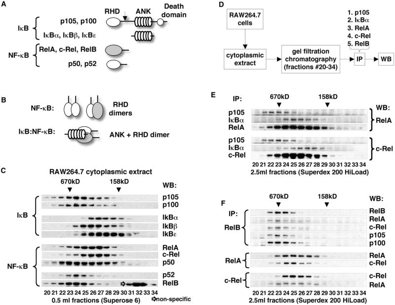
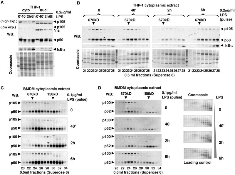
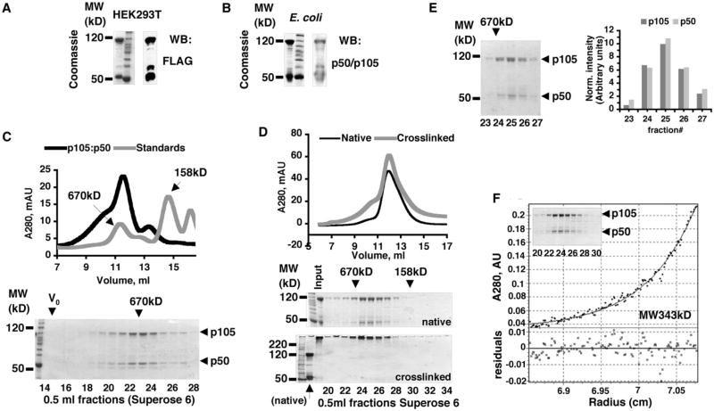
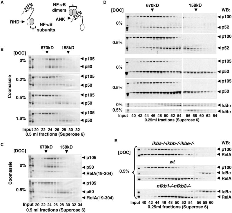
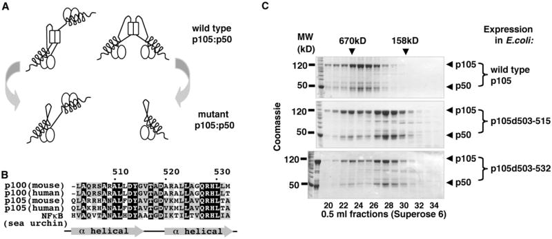
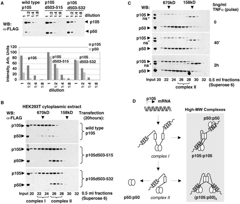
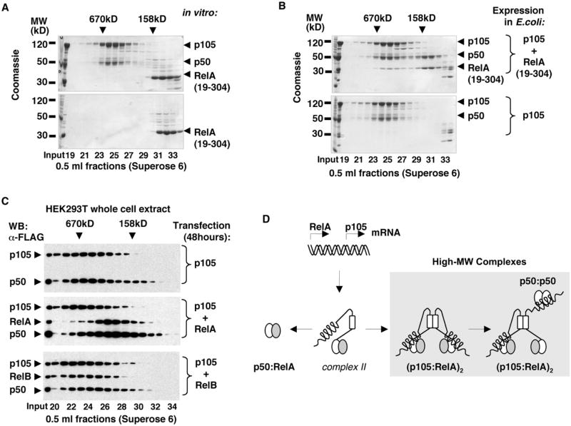
Nfkb1 and Nfkb2 proteins p105 and p100 serve both as NF-kappaB precursors and inhibitors of NF-kappaB dimers. In a biochemical characterization of endogenous cytoplasmic and purified recombinant proteins, we found that p105 and p100 assemble into high-molecular-weight complexes that contribute to the regulation of all NF-kappaB isoforms. Unlike the classical inhibitors IkappaBalpha, -beta, and -epsilon, high-molecular-weight complexes of p105 and p100 proteins bind NF-kappaB subunits in two modes: through direct dimerization of Rel homology domain-containing NF-kappaB polypeptides and through interactions of the p105 and p100 ankyrin repeats with preformed NF-kappaB dimers, thereby mediating the bona fide IkappaB activities, IkappaBgamma and IkappaBdelta. Our biochemical evidence suggests an assembly pathway in which kinetic mechanisms control NF-kappaB dimer formation via processing and assembly of large complexes that contain IkappaB activities.


Figures

Figure 1
Figure 1. Detection of Endogenous High-MW p105 and p100 Complexes
(A) Domain organization of IκB and NF-κB proteins. RHD, Rel homology domain; ANK, ankyrin repeat domain. Arrow indicates site of proteolysis in p105 and p100 that yields p50 and p52, respectively. (B) Domain interactions between NF-κB and IκB proteins. Dimerization of RHD domains (top) and binding of ANK domain to RHD dimers (bottom). (C) Cytoplasmic extract of RAW264.7 cells was fractionated by gel filtration chromatography (GF) on an analytical Superose 6 column. IκB and NF-κB proteins were detected by western blotting (WB). (D) Flowchart illustrating immunoprecipitation (IP) experiments shown in (E) and (F). (E) Cytoplasmic extract of RAW264.7 cells was fractionated by GF. p105, IκBα, RelA, and c-Rel were immunoprecipitated from GF fractions. RelA (top) and c-Rel (bottom) were detected by WB. (F) RelB, RelA, and c-Rel were immunoprecipitated from GF fractions (as in E). RelB, RelA, c-Rel, p105, and p100 were detected by WB.
Figure 2
Figure 2. High-MW Endogenous p105 and p100 Complexes Are Stimulus Responsive
(A) THP-1 cells were stimulated with 0.2 μg/ml LPS for 40 min. Cytoplasmic (cyto) and nuclear (nucl) extracts were prepared at indicated time points and analyzed by western blotting (WB) to detect p105, p50, and IκBα. Exp., exposure. (B) Cytoplasmic extracts of THP-1 cells (as in A) were further fractionated by gel filtration chromatography (GF) and analyzed by WB to detect p105, p50, and IκBα. (C) BMDM were stimulated with 0.1 μg/ml LPS for 40 min. Cytoplasmic extracts were prepared at indicated time points and analyzed by GF followed by WB to detect p105 and p50 proteins. (D) GF fractions of BMDM (as shown in C) were analyzed by WB to detect p100 and p52. Coomassie staining (in parallel with WB) provided controls for equivalent loading and for the efficiency of fractionation.
Figure 3
Figure 3. Stoichiometry of p105:p50 Complexes
(A and B) p105 was purified by affinity chromatography from the mammalian (HEK293T, A) and bacterial (E. coli, B) expression systems. p105 and copurified 50 kD N-terminal fragment of p105 were detected by Coomassie staining and western blotting (WB). (C) p105:p50 (purified from HEK293T) was analyzed by gel filtration chromatography (GF) and compared to MW standards. Absorbance at λ280 nm (A280) was plotted against retention volume (top). The resulting fractions were analyzed by SDS-PAGE and Coomassie staining (bottom). (D) Chemically crosslinked p105:p50 complexes and native p105:p50 complexes were compared by GF on an analytical Superose 6 column. A280 was plotted against retention volume (top). The resulting fractions and input samples were analyzed by SDS-PAGE and Coomassie staining (bottom). (E) 100 μg of p105:p50 complex was fractionated by GF followed by SDS-PAGE and Coomassie staining (left). The normalized values of the intensities of p105 and p50 bands were plotted against fraction number (right). (F) p105:p50 complexes were purified from E. coli by Ni affinity chromatography followed by GF (insert, Coomassie-stained SDS-PAGE gel shows GF fractions) and analyzed by analytical ultracentrifugation (AUC). The representative result of the sedimentation equilibrium (SE) experiment performed at the rotor speed 8000 rpm is shown.
Figure 4
Figure 4. Two Modes of Binding of NF-κB Subunits to p105 and p100
(A) Individual NF-κB subunits interact with p105 and p100 via dimerization of RHD domains (left); preformed NF-κB dimers interact with p105 or p100 ANK domains (right). (B) Recombinant p105:p50 complexes were analyzed by gel filtration chromatography (GF) in the presence of 0%-1.6% of sodium deoxycholate (DOC) followed by SDS-PAGE and Coomassie staining. (C) Recombinant p105:p50:RelA(19–304) complexes were analyzed by GF in standard conditions and in the presence of 0.8% DOC followed by SDS-PAGE and Coomassie staining. (D) Cytoplasmic extracts of THP-1 cells were analyzed by GF in standard conditions and in the presence of 0.5% DOC followed by western blotting (WB) to detect p100 and p52 (top), p105 and p50 (middle), or IκBα (bottom). (E) Cytoplasmic extracts of ikba−/−ikb−/−ikbbe−/−, wild-type (WT), and nfkb1−/−nfkb2−/− MEF were analyzed by GF in the presence of 0.5% DOC. p100, IκBα, and RelA were detected by WB.
Figure 5
Figure 5. Molecular Architecture of p105:p50 Complexes
(A) The predicted architectures of the minimal (core) wild-type p105:p50 complexes (top) and complexes formed by the C-terminal helical dimerization domain mutants of p105 (bottom). (B) A multiple sequence alignment of the predicted α-helical dimerization domain of human and mouse p105, p100, and their ortholog (NFkB) from sea urchin. (C) Wild-type and mutant p105 proteins, p105d503–515 and p105d503–532, were expressed in E.coli and purified by the Ni affinity chromatography (via N-terminal His tag) followed by gel filtration chromatography (GF). GF fractions were analyzed by SDS-PAGE and Coomassie staining.
Figure 6
Figure 6. p105 Processing and the Assembly of p105 High-MW Complexes
(A) FLAG-tagged wild-type (WT) and mutant p105 proteins, p105d503–515 and p105d503–532, were transiently expressed in HEK293T cells. Cytoplasmic extracts were prepared 20 hr after transfection, and their serial dilutions were analyzed by western blotting (WB, top). The intensities of WT and mutant p105 and p50 bands were quantitated and plotted against dilution factor (bottom). (B) Cytoplasmic extracts (as in A)were analyzed by gel filtration chromatography (GF) followed by WB. Complex I and complex II denote p105 complexes distinct in their chromatographic properties. (C) HEK293T cells were stimulated with 5 ng/ml TNFα for 40 min. Cytoplasmic extracts were prepared at indicated time points and analyzed by GF followed by WB to detect endogenous p105 and p50 proteins. Complex II denotes the distinct lower-MW p105 complexes detected after stimulation. (D) A model for the biogenesis of p50 homodimers and the high-MW p105:p50 complexes.
Figure 7
Figure 7. Biogenesis of p50:RelA Heterodimers and RelA Complexes with p105
(A) Recombinant p105:p50 complexes were incubated with RelA RHD homodimers, RelA(19–304), and the resulting complexes were analyzed by gel filtration chromatography (GF) followed by SDS-PAGE and Coomassie staining (top) and compared to the similarly treated isolated RelA(19–304) (bottom). (B) His-tagged p105 was coexpressed with untagged RelA(19–304) in E. coli, and the resulting complexes were isolated by Ni affinity chromatography followed by GF (top) and compared to the recombinant p105:p50 complexes (bottom). (C) FLAG-tagged p105 was expressed in HEK293T cells alone or coexpressed with FLAG-tagged RelA or RelB. At 48 hr after transfection, the whole-cell extracts were analyzed by GF followed by western blotting (WB). (D) A model for the biogenesis of p50:RelA heterodimers and the high-MW p105:p50:RelA complexes.

References

    1. Baeuerle PA, Baltimore D. Activation of DNA-binding activity in an apparently cytoplasmic precursor of the NF-kappa B transcription factor. Cell. 1988;53:211–217. - PubMed
    1. Basak S, Kim H, Kearns JD, Tergaonkar V, O'Dea E, Werner SL, Benedict CA, Ware CF, Ghosh G, Verma IM, Hoffmann A. A fourth IkappaB protein within the NF-kappaB signaling module. Cell. 2007;128:369–381. - PMC - PubMed
    1. Beinke S, Deka J, Lang V, Belich MP, Walker PA, Howell S, Smerdon SJ, Gamblin SJ, Ley SC. NF-kappaB1 p105 negatively regulates TPL-2 MEK kinase activity. Mol Cell Biol. 2003;23:4739–4752. - PMC - PubMed
    1. Caamano JH, Rizzo CA, Durham SK, Barton DS, Raventos-Suarez C, Snapper CM, Bravo R. Nuclear factor (NF)-kappa B2 (p100/p52) is required for normal splenic microarchitecture and B cell-mediated immune responses. J Exp Med. 1998;187:185–196. - PMC - PubMed
    1. Compton SJ, Jones CG. Mechanism of dye response and interference in the Bradford protein assay. Anal Biochem. 1985;151:369–374. - PubMed

Publication types

MeSH terms