Comparative Study
. 2009 Oct;70(10):790-802.
doi: 10.1016/j.humimm.2009.06.005. Epub 2009 Jun 12.

Comparison of human fetal liver, umbilical cord blood, and adult blood hematopoietic stem cell engraftment in NOD-scid/gammac-/-, Balb/c-Rag1-/-gammac-/-, and C.B-17-scid/bg immunodeficient mice

Affiliations
Comparative Study

Comparison of human fetal liver, umbilical cord blood, and adult blood hematopoietic stem cell engraftment in NOD-scid/gammac-/-, Balb/c-Rag1-/-gammac-/-, and C.B-17-scid/bg immunodeficient mice

Christin M Lepus et al. Hum Immunol. 2009 Oct.

Abstract

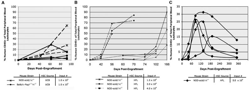
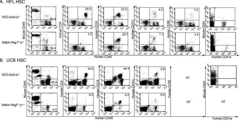
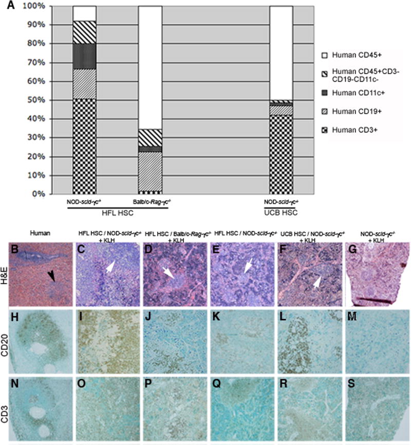
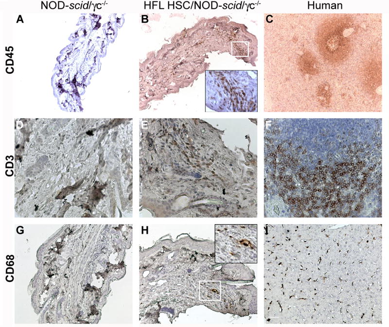
Immunodeficient mice bearing components of a human immune system present a novel approach for studying human immune responses. We investigated the number, phenotype, developmental kinetics, and function of developing human immune cells following transfer of CD34(+) hematopoietic stem cell (HSC) preparations originating from second trimester human fetal liver (HFL), umbilical cord blood (UCB), or granulocyte colony-stimulating factor-mobilized adult blood (G-CSF-AB) delivered via intrahepatic injection into sublethally irradiated neonatal NOD-scid/gammac(-/-), Balb/c-Rag1(-/-)gammac(-/-), and C.B-17-scid/bg mice. HFL and UCB HSC provided the greatest number and breadth of developing cells. NOD-scid/gammac(-/-) and Balb/c-Rag1(-/-)gammac(-/-) harbored human B and dendritic cells as well as human platelets in peripheral blood, whereas NOD-scid/gammac(-/-) mice harbored higher levels of human T cells. NOD-scid/gammac(-/-) mice engrafted with HFL CD34(+) HSC demonstrated human immunological competence evidenced by white pulp expansion and increases in total human immunoglobulin following immunization with T-dependent antigens and delayed-type hypersensitivity-infiltrating leukocytes in response to antigenic challenge. In conclusion, we describe an encouraging base system for studying human hematopoietic lineage development and function utilizing human HFL or UCB HSC-engrafted NOD-scid/gammac(-/-) mice that is well suited for future studies toward the development of a fully competent humanized mouse model.


Figures

Figure 1
Figure 1. Kinetics of human CD45+ cell development in NOD-scid/γc−/− and Balb/c-Rag1−/−γc−/−mice injected as neonates
Prior to 2 days of age, NOD-scid/γc−/− or Balb/c-Rag1−/−γc−/− mice were irradiated and then administered HFL or UCB HSC preparations by intrahepatic injection. The percentage of human CD45+ cells of the total leukocyte population in peripheral blood was determined in successive post-engraftment dates by flow cytometry in various cohorts of mice. A: 3 month comparative analysis of individual NOD-scid/γc−/− or Balb/c-Rag1−/−γc−/− receiving 1.0 × 10⁵ UCB HSC; B: 6 month comparative analysis of individual NOD-scid/γc−/− mice receiving either 1.5 × 10⁴, 3.0 × 10⁴ or 4.0 × 10⁴ HFL HSC; C: 12 month comparative analysis of individual NOD-scid/γc−/− mice receiving 5.0 × 10⁴ HFL HSC.
Figure 2
Figure 2. Development of human immune cell subsets in NOD-scid/γc−/− and Balb/c-Rag1−/−γc−/− mice receiving HFL HSC or UCB HSC
At 1 day of age, NOD-scid/γc−/− or Balb/c-Rag1−/−γc−/− mice were irradiated and then injected intrahepatically with 4.0 × 10⁴ HFL HSC (A) or 1.0 × 10⁵ UCB HSC (B). The phenotype of human CD45+ cells expressing CD3, CD19, CD11c, or CD56 (at 8 or 13 weeks of age in A or B, respectively) as well as CD41a+ platelets (at 15 weeks or 28 weeks of age in A or B, respectively), in peripheral blood from HFL HSC- or UCB HSC-engrafted mice was determined by flow cytometry. Representative sets of plots from a NOD-scid/γc−/− and a Balb/c-Rag1−/−γc−/− mouse are shown in the top and bottom row of panel A (HFL HSC-engrafted mice) and panel B (UCB HSC-engrafted mice), respectively. NT = not tested.
Figure 3
Figure 3. Dependence of human T and B lymphocyte development on HFL HSC inocula number in NOD-scid/γc−/− mice
At 1 day of age, NOD-scid/γc−/− mice were irradiated and then injected intrahepatically with 1.5 – 5.0 × 10⁴ HFL HSC. A: Flow cytometric plots showing human CD45+CD3+ cells in peripheral blood at 10 weeks of age from individual mice injected with either 1.5 × 10⁴ or 3.0 × 10⁴ HFL HSC. B: Percentages of CD45+CD3+ and CD45+CD19+ cells of total lymphocytes determined by flow cytometric analysis of peripheral blood at 10 – 12 weeks of age from individual mice injected with increasing numbers of HFL HSC.
Figure 4
Figure 4. Analysis of human immune cell subsets in spleens of representative NOD-scid/γc−/− and Balb/c-Rag1−/−γc−/− mice receiving HFL or UCB HSC
At 1 day of age, NOD-scid/γc−/− or Balb/c-Rag1−/−γc−/− mice were irradiated and then injected intrahepatically with 4.0 × 10⁴ HFL HSC or 1.0 × 10⁵ UCB HSC or left as non-irradiated, non-transplanted controls. The phenotype of human CD45+ splenocytes (CD3, CD19, or CD11c) from representative HFL HSC- or UCB HSC-engrafted mice was determined by flow cytometry following euthanasia at 15 or 28 weeks of life, respectively (A). Paraffin-embedded sections originating from a human spleen (B, H, N), or spleens from a KLH-immunized, HFL HSC-engrafted NOD-scid/γc−/− mouse (C, I, O), KLH-immunized, HFL HSC-engrafted Balb/c-Rag1−/− γc−/− mouse (D, J, P), non-immunized, HFL HSC-engrafted NOD-scid/γc−/− mouse (E, K, Q), KLH-immunized, UCB HSC-engrafted NOD-scid/γc−/− mouse (F, L, R), and KLH-immunized, non-engrafted NOD-scid/γc−/− mouse (G, M, S) were stained with H&E, CD20, or CD3, respectively (H&E: 4x magnification, CD20 and CD3: 10x magnification). White pulp in human splenic section and white pulp-like regions in engrafted mice are shown in panel B (black arrow) and panel C – F (white arrows), respectively.
Figure 5
Figure 5. Analysis of thymi and lymph nodes in NOD-scid/γc−/− and Balb/c-Rag1−/−γc−/− mice receiving HFL or UCB HSC
A – C: Paraffin-embedded, H&E-stained thymic tissue sections from an immunocompetent NOD mouse (A, 4x); NOD-scid/γc−/− (B, 4x) and Balb/c-Rag1−/−γc−/− (C, 10x) mice engrafted 28 weeks earlier with 4.0 × 10⁴ HFL HSC. White and red arrows show medullary-like and cortical-like regions, respectively. D – F: H&E-stained thymic tissue section (D, 4x) and flow cytometric plots of thymic leukocytes showing human CD45+CD3+ cells (E) and CD4+, CD8+ and CD4+CD8+ cells (F) from representative NOD-scid/γc−/− mice injected with 1.0 × 10⁵ UCB HSC and analyzed at 28 weeks of age. G – I: Paraffin-embedded mesenteric (G, H; 4x and 20x magnification, respectively) and popliteal (I, 40x magnification) lymph node sections were harvested at 12 months of age from influenza-immunized NOD-scid/γc−/− mice that received 5.0 × 10⁴ HFL HSC as neonates and stained with either H&E or anti-human CD45 (insets).
Figure 6
Figure 6. Splenocyte blastogenesis and human-specific immunoglobulin responses in HSC-engrafted, KLH- or influenza-immunized NOD-scid/γc−/− and Balb/c-Rag1−/− γc−/− mice
At 1 day of age, NOD-scid/γc−/− and Balb/c-Rag1−/−γc−/− mice were irradiated and then injected intrahepatically with 4.0 × 10⁴ HFL HSC (KLH-immunized group), 5.0 × 10⁴ HFL HSC (influenza-immunized group), 1.0 × 10⁵ UCB HSC (KLH-immunized group), or left as non-irradiated, non-transplanted controls. A: At 15 weeks of age, splenocytes were harvested and cultured in the presence or absence of 2 μg/ml phytohemagglutinin (PHA). An 18 hour 3H-thymidine incorporation pulse was determined in triplicate beginning at 48 hours of culture (+/− standard deviation). * indicates HSC-engrafted mice with <1% peripheral blood chimerism. B-D: Mice began a series of 2 intradermal immunization doses with either KLH at 12 weeks (HFL HSC-engrafted) or 25 weeks (UCB HSC-engrafted) or influenza at 21 weeks. Total human IgM, total human IgG, and influenza-specific human (IgM+IgG+IgA) immunoglobulin (B-D, respectively), present in the chimeric mouse plasma, both immediately prior to the first immunization dose (pre) and 2 weeks following the second dose (post), was determined by ELISA. E – F: Flow cytometric plots showing human CD19+ and CD5+ co-expression (F) in the gated human CD45+ population (E) in peripheral blood leukocytes from a representative Balb/c-Rag−/−γc−/− mouse injected at 2 days of age with 1.0 × 10⁵ UCB HSC and analyzed at 11 weeks of age.
Figure 7
Figure 7. Delayed type hypersensitivity responses in HFL HSC-engrafted, KLH-immunized NOD-scid/γc−/− mice
At 1 day of age, NOD-scid/γc−/− mice were irradiated and then injected intrahepatically with 4.0 × 10⁴ HFL HSC or left as an non-irradiated, non-transplanted controls. Mice were immunized twice intradermally with KLH then challenged in the footpad and the ear 18 days after their initial KLH exposure. Paraffin-embedded footpad sections originating from a challenged, non-engrafted (A, D, G) or engrafted (B, E, H) KLH-immunized NOD-scid/γc−/− mouse, harvested at 15 weeks of age, or from relevant human sections were stained for human CD45 (A – C; 10x magnification), human CD3 (D-F; 40x magnification), or human CD68 (G-I; 20x magnification). Insets in B and H are 40x magnifications of the respective white-boxed regions.

References

    1. Melkus MW, Estes JD, Padgett-Thomas A, Gatlin J, Denton PW, Othieno FA, et al. Humanized mice mount specific adaptive and innate immune responses to EBV and TSST-1. Nat Med. 2006 Nov;12(11):1316–1322. - PubMed
    1. Shultz LD, Ishikawa F, Greiner DL. Humanized mice in translational biomedical research. Nat Rev Immunol. 2007 Feb;7(2):118–130. - PubMed
    1. Zollner TM, Podda M, Pien C, Elliott PJ, Kaufmann R, Boehncke WH. Proteasome inhibition reduces superantigen-mediated T cell activation and the severity of psoriasis in a SCID-hu model. J Clin Invest. 2002 Mar;109(5):671–679. - PMC - PubMed
    1. Pober JS, Bothwell AL, Lorber MI, McNiff JM, Schechner JS, Tellides G. Immunopathology of human T cell responses to skin, artery and endothelial cell grafts in the human peripheral blood lymphocyte/severe combined immunodeficient mouse. Springer Semin Immunopathol. 2003 Sep;25(2):167–80. - PubMed
    1. Banuelos SJ, Shultz LD, Greiner DL, Burzenski LM, Gott B, Lyons BL, et al. Rejection of human islets and human HLA-A2.1 transgenic mouse islets by alloreactive human lymphocytes in immunodeficient NOD-scid and NOD-Rag1(null)Prf1(null) mice. Clin Immunol. 2004 Sep;112(3):273–283. - PubMed

Publication types

MeSH terms