. 2009 Oct;23(10):3393-404.
doi: 10.1096/fj.09-135467. Epub 2009 Jun 12.

Genetic requirements for Staphylococcus aureus abscess formation and persistence in host tissues

Affiliations

Genetic requirements for Staphylococcus aureus abscess formation and persistence in host tissues

Alice G Cheng et al. FASEB J. 2009 Oct.

Erratum in

  • FASEB J. 2010 Feb;24(2):648

Abstract

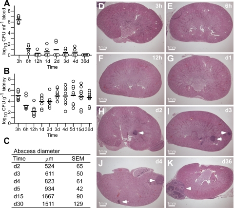
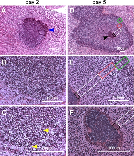
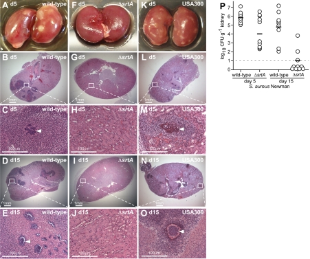
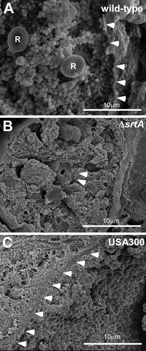
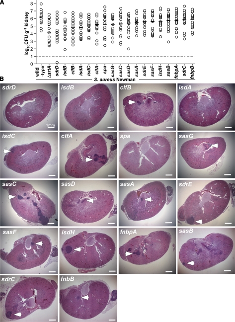
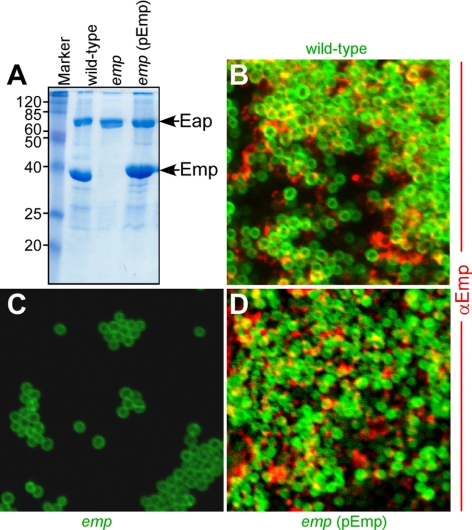
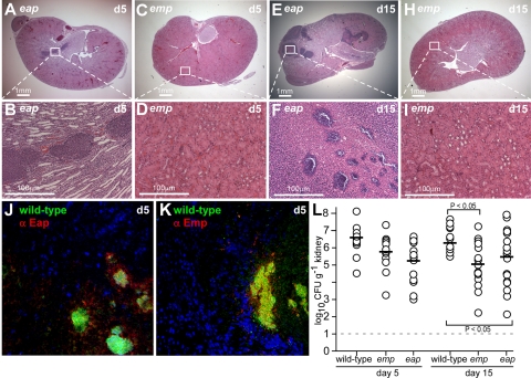
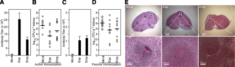
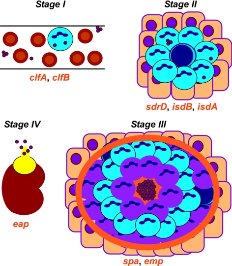
Staphylococcus aureus infections are associated with abscess formation and bacterial persistence; however, the genes that enable this lifestyle are not known. We show here that following intravenous infection of mice, S. aureus disseminates rapidly into organ tissues and elicits abscess lesions that develop over weeks but cannot be cleared by the host. Staphylococci grow as communities at the center of abscess lesions and are enclosed by pseudocapsules, separating the pathogen from immune cells. By testing insertional variants in genes for cell wall-anchored surface proteins, we are able to infer the stage at which these molecules function. Fibrinogen-binding proteins ClfA and ClfB are required during the early phase of staphylococcal dissemination. The heme scavenging factors IsdA and IsdB, as well as SdrD and protein A, are necessary for abscess formation. Envelope-associated proteins, Emp and Eap, are either required for abscess formation or contribute to persistence. Fluorescence microscopy revealed Eap deposition within the pseudocapsule, whereas Emp was localized within staphylococcal abscess communities. Antibodies directed against envelope-associated proteins generated vaccine protection against staphylococcal abscess formation. Thus, staphylococci employ envelope proteins at discrete stages of a developmental program that enables abscess formation and bacterial persistence in host tissues.


Figures

Figure 1.
Figure 1.
Staphylococcal abscess formation following intravenous infection of mice. A) BALB/c mice were infected with 1 × 10⁷ CFU of S. aureus Newman by retro-orbital injection. Cohorts of 5 mice were examined by cardiac puncture at timed intervals for bacterial load in blood; sample aliquots were plated on agar medium, and colony formation was enumerated (CFU/ml blood). Black bars indicate means of observations. B) Dissemination of S. aureus Newman into peripheral organ tissues and replication of the pathogen was measured at timed intervals in the kidneys of mice (cohorts of 10 animals), which were homogenized and plated on agar medium for colony formation and enumeration. C) Diameter of abscess lesions was measured in thin-sectioned H&E-stained tissues of infected kidneys at timed intervals. D–K) Images of infected kidneys at timed intervals analyzed in thin-sectioned H&E-stained tissues. Arrowheads indicate abscess lesions.
Figure 2.
Figure 2.
Histopathology of SACs. BALB/c mice were infected with S. aureus Newman via retro-orbital injection. Thin-sectioned, H&E-stained tissues of infected kidneys on d 2 (A–C) and d 5 following infection (D–F) were analyzed by light microscopy, and images were captured. On d 2, a massive infiltrate (A, blue arrowhead) of PMNs with occasional intracellular staphylococci (C, yellow arrowheads) are characteristic of early infectious lesions. By d 5, staphylococcal abscess communities developed as a central nidus (D, black arrowhead). Staphylococci were enclosed by an amorphous, eosinophilic pseudocapsule (black box) and surrounded by a zone of dead PMNs (white box), a zone of apparently healthy PMNs (red box), and a rim of necrotic PMNs (green box), separated through an eosinophilic layer from healthy kidney tissue.
Figure 3.
Figure 3.
Sortase A is required for abscess formation and staphylococcal persistence in host tissues. Kidneys of BALB/c mice (cohorts of 10 animals) infected with S. aureus Newman, its isogenic sortase A mutant (ΔsrtA), or methicillin-resistant S. aureus USA300 were removed during necropsy of animals at d 5 and 15 following inoculation. A–O) Kidneys were inspected for surface abscesses (A, F, K) or fixed in formalin, embedded, thin sectioned, and stained with H&E. Histopathology images were acquired with light microscopy at ×12.5 (B, G, L, D, I, N) and ×200 (C, H, M, E, J, O). P) Staphylococcal replication and persistence in kidney tissue was measured at d 5 and 15 following infection. Kidneys were removed from infected mice during necropsy; tissue was homogenized and plated on agar medium for colony formation and enumeration.
Figure 4.
Figure 4.
Staphylococcal communities at the center of abscess lesions. Kidney tissue from mice infected with S. aureus Newman (wild type), its isogenic sortase A mutant (ΔsrtA), or MRSA strain USA300 was sectioned, fixed, dehydrated, and sputter-coated with 80% Pt/20% Pd for scanning electron microscopy. A) Wild-type pathogen is organized as a tightly associated lawn, the SAC, at the abscess center that is contained within an amorphous pseudocapsule (arrowheads), separating SACs from the cuff of leukocytes. Red blood cells (R) are located among staphylococci. B) The sortase mutant, ΔsrtA (arrowheads) did not form SACs, and isolated staphylococci were found in healthy kidney tissue. C) Similar to S. aureus Newman, MRSA strain USA300 also formed SACs contained within a pseudocapsule (arrowheads).
Figure 5.
Figure 5.
Formation of SACs requires specific surface proteins. A) S. aureus Newman variants with bursa aurealis insertions in surface-protein genes were examined 5 d following infection of BALB/c mice (cohorts of 20 animals) for bacterial load in homogenized kidney tissues. B) H&E-stained thin sections of infected kidneys were examined by light microscopy at ×12.5 for abscess lesions (arrowheads).
Figure 6.
Figure 6.
The envelope-associated protein Emp is a component of the extracellular matrix in staphylococcal biofilms. A) Coomassie-stained SDS-PAGE identified envelope-associated proteins Eap and Emp and characterized a bursa aurealis mutant (emp) with (pEmp) and without complementing plasmid. BD) Expression of Emp in the extracellular matrix of in vitro-formed biofilms was detected by microscopy, using BODIPY-vancomycin-stained staphylococci (green) and AlexaFluor-647-labeled secondary antibody to recognize the deposition of rabbit anti-Emp (red).
Figure 7.
Figure 7.
Emp and Eap in staphylococcal abscess lesions. Kidneys of BALB/c mice infected with S. aureus Newman variants carrying bursa aurealis insertions in emp or eap were removed at d 5 and 15 following inoculation. A–K) Kidneys were stained with H&E, and histopathology images were acquired with light microscopy at ×12.5 (A, C, E, H) and ×200 (B, D, F, I). Expression of Eap (J) and Emp (K) in abscess lesions of wild-type S. aureus Newman was detected with rabbit anti-Emp or anti-Eap and secondary AlexaFluor-647 labeled antibodies (red) in renal tissue stained with Hoechst dye (blue) to detect nuclei of polymorphonuclear leukocytes, and with BODIPY-vancomycin (green) to reveal staphylococcal abscess communities. L) Staphylococcal replication and persistence in kidney tissue was measured at d 5, 15, and 30 following intravenous inoculation. Kidneys were removed from infected mice (cohorts of 10 animals), and tissue was homogenized and plated on agar medium for colony formation and enumeration.
Figure 8.
Figure 8.
Active and passive immunization with Eap generates protection from staphylococcal challenge. A) BALB/c mice were immunized with purified Eap or Emp or mock treated with adjuvant alone, and serum IgG titers were analyzed by ELISA. B) Three weeks following immunization, animals were challenged via intravenous inoculation of staphylococci. Five days following infection, kidneys were removed during necropsy, and renal tissue was analyzed for staphylococcal load or histopathology. C) Rabbit antibodies directed against Eap or Emp were purified by affinity chromatography and passively transferred by intraperitoneal injection into mice. Twenty-four hours later, serum IgG titers of passively immunized animals were analyzed by ELISA. D) Animals passively immunized with purified antibodies against Eap or Emp, as well as mock-immunized animals were subsequently challenged with S. aureus Newman, and bacterial load was enumerated on d 4. E) Abscess formation in kidneys was detected in thin-sectioned, H&E-stained tissues.
Figure 9.
Figure 9.
A working model for staphylococcal abscess formation and persistence in host tissues. Stage I: following intravenous inoculation, S. aureus survives in the bloodstream and disseminates via the vasculature to peripheral organ tissues. Stage II: in renal tissues, staphylococci attract a massive infiltrate of polymorphonuclear leukocytes and other immune cells. Stage III: abscesses mature with a central accumulation of the pathogen (SAC), enclosed by an eosinophilic pseudocapsule. The SAC is surrounded by a zone of dead PMNs, apparently healthy PMNs, and finally an outer zone of dead PMNs with a rim of eosinophilic material. Stage 4: abscesses mature and rupture on the organ surface, thereby releasing staphylococci into circulation and initiating new rounds of abscess development. Genes for bacterial envelope components that are required for specific stages of staphylococcal abscess development are in red below the corresponding stage during which these genes function. See text for details.

References

    1. Gorbach S L. Good and laudable pus. J Clin Invest. 1995;96:2545. - PMC - PubMed
    1. Tzianabos A O, Kasper D L, Cisneros R L, Smith R S, Onderdonk A B. Polysaccharide-mediated protection against abscess formation in experimental intra-abdominal sepsis. J Clin Invest. 1995;96:2727–2731. - PMC - PubMed
    1. Wallenfang T, Bohl J, Kretzschmar K. Evolution of brain abscess in cats formation of capsule and resolution of brain edema. Neurosurg Rev. 1980;3:101–111. - PubMed
    1. Russell D G. Staphylococcus and the healing power of pus. Cell Host Microbe. 2008;3:115–116. - PubMed
    1. Klevens R M, Morrison M A, Nadle J, Petit S, Gershman K, Ray S, Harrison L H, Lynfield R, Dumyati G, Townes J M, Craig A S, Zell E R, Fosheim G E, McDougal L K, Carey R B, Fridkin S K, Investigators for the Active Bacterial Core surveillance (ABCs) MRSA Investigators Invasive methicillin-resistant Staphylococcus aureus infections in the United States. JAMA. 2007;298:1763–1771. - PubMed

Publication types

Substances