. 2009 Sep;8(9):2170-85.
doi: 10.1074/mcp.M900088-MCP200. Epub 2009 Jun 12.

Identification and quantification of glycoproteins using ion-pairing normal-phase liquid chromatography and mass spectrometry

Affiliations

Identification and quantification of glycoproteins using ion-pairing normal-phase liquid chromatography and mass spectrometry

Wen Ding et al. Mol Cell Proteomics. 2009 Sep.

Abstract

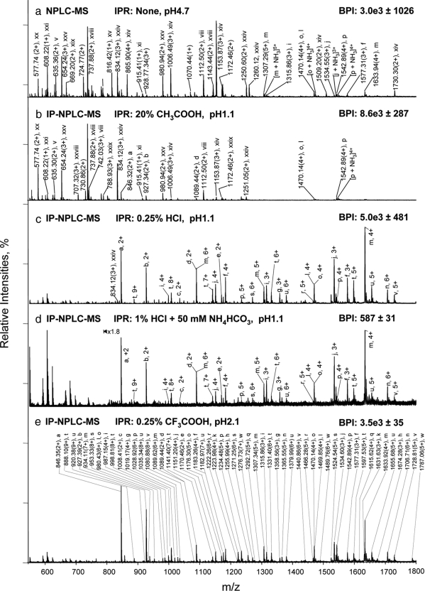
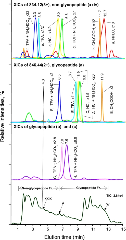
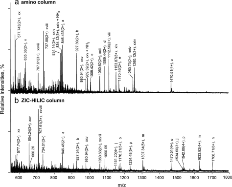
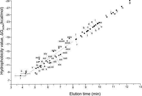
Glycoprotein structure determination and quantification by MS requires efficient isolation of glycopeptides from a proteolytic digest of complex protein mixtures. Here we describe that the use of acids as ion-pairing reagents in normal-phase chromatography (IP-NPLC) considerably increases the hydrophobicity differences between non-glycopeptides and glycopeptides, thereby resulting in the reproducible isolation of N-linked high mannose type and sialylated glycopeptides from the tryptic digest of a ribonuclease B and fetuin mixture. The elution order of non-glycopeptides relative to glycopeptides in IP-NPLC is predictable by their hydrophobicity values calculated using the Wimley-White water/octanol hydrophobicity scale. O-linked glycopeptides can be efficiently isolated from fetuin tryptic digests using IP-NPLC when N-glycans are first removed with PNGase. IP-NPLC recovers close to 100% of bacterial N-linked glycopeptides modified with non-sialylated heptasaccharides from tryptic digests of periplasmic protein extracts from Campylobacter jejuni 11168 and its pglD mutant. Label-free nano-flow reversed-phase LC-MS is used for quantification of differentially expressed glycopeptides from the C. jejuni wild-type and pglD mutant followed by identification of these glycoproteins using multiple stage tandem MS. This method further confirms the acetyltransferase activity of PglD and demonstrates for the first time that heptasaccharides containing monoacetylated bacillosamine are transferred to proteins in both the wild-type and mutant strains. We believe that IP-NPLC will be a useful tool for quantitative glycoproteomics.


Figures

Fig. 1.
Fig. 1.
Isolation of glycopeptides from a RNase B and fetuin tryptic digest using IP-NPLC. Total ion mass spectrum from NPLC-ESI-MS of peptides in the glycopeptide fraction from the tryptic digest injected with the following ion-pairing reagents (IPR): a, none in 80% ACN/20% H2O; (pH 4.7); b, 20% acetic acid in 80% ACN (pH 1.1); c, 0.25% HCl in 80% ACN/20% H2O (pH 1.1); d, 50 mm NH4HCO3 + 1% HCl in 80% ACN/20% H2O (pH 1.1); and e, 0.25% TFA in 85% ACN/15% H2O (pH 2.1). BPI, base peak intensities. Throughout this report, the total ion mass spectrum for NPLC-ESI-MS of the glycopeptide fraction refers to the summed MS scans across the NPLC retention times between the most hydrophobic high mannose type glycopeptide (a) from RNase B (6.5–9.0 min depending on the ion-pairing reagents used (Fig. 2, 2nd top panel) and the most hydrophilic sialylated glycopeptide from fetuin detected (earlier than 13 min). Non-glycopeptides eluted earlier than the high mannose type glycopeptide (a) of RNase B when HCl or TFA were used as ion-pairing reagents. The tryptic digest contains 14.2 pmol of RNase B tryptic digest + 13.8 pmol of fetuin tryptic digest. The solvent compositions used to dissolve the different IPR, CH3COOH, HCl, and TFA with/without NH4HCO3 are as indicated in each panel of the figure. The pH values listed are those of the samples. All abundant ions that are not labeled in Fig. 1 are singly charged contaminants.
Fig. 2.
Fig. 2.
Effect of acids as ion-pairing reagents on peptide retention in IP-NPLC. Top panel, overlaid extracted ion chromatograms (XICs) of m/z 834.12 (3+) of the hydrophilic non-glycopeptide (xxiv), CKPVNTFVHESLADVQAVCSQK, from the RNase B and fetuin tryptic digest injected with the following ion-pairing reagents (IPR): (a) none; (b) 20% CH3COOH; (c) 0.25% HCl; (d) 1% HCl + 50 mm NH4HCO3; (e) 0.25% TFA; and (f) 1% TFA + 50 mm NH4HCO3. Second top panel, overlaid XICs of m/z 846.44 (2+) of the least hydrophilic glycopeptide (a), NLTK-GlcNAc2Man5, from the same tryptic digest injected with the following IPRs: (B) 20% CH3COOH; (C) 0.25% HCl; (D) 1%HCl + 50 mm NH4HCO3; (E) 0.25% TFA; and (F) 1% TFA + 50 mm NH4HCO3; Second bottom panel, (G) XIC of m/z 927.48 (2+) of the glycopeptide (b), NLTK-GlcNAc2Man6; (H) XIC of m/z 1008.43 (2+) of the glycopeptide (c), NLTK-GlcNAc2Man7. IPR used for G and H: 1% TFA + 50 mm NH4HCO3. Bottom panel, total ion chromatogram from IP-NPLC-ESI-MS of the standard tryptic digest injected with 1% TFA + 50 mm NH4HCO3. The magnification factors (e.g. x1) of the base peak intensity (BPI) of the extracted ions were relative to the BPI of (E) for normalization. The elution time of the least hydrophilic tryptic glycopeptide (a) of RNase B, i.e. the reference time determining the starting elution time of glycopeptide fraction, varies from 6.5 min to 11.9 min depending on the IPR used (Second top panel). The broken line at 4.3 min indicates the elution time of the most hydrophilic tryptic non-glycopeptide (xxix) present in the sample. w, glycopeptide (w) in Table II; Fr., fraction.
Fig. 3.
Fig. 3.
Effect of stationary phase charges on glycopeptide isolation using IP-NPLC. Total ion mass spectrum from NPLC-ESI-MS of peptides in the glycopeptide fraction from the RNase B and fetuin tryptic digest injected with 1% TFA + 50 mm NH4HCO3 using different hydrophilic columns: (a) amino; (b) ZIC-HILIC. All other abundant ions that are not labeled in Fig. 3 are singly charged contaminants. Ammonium adducts and minor peptide peaks are not labeled.
Fig. 4.
Fig. 4.
Plot of the hydrophobicity values of the non-glycopeptides of the RNase B and fetuin tryptic digest versus their elution times in IP-NPLC. The sample was injected with 0.25% HCl (pH 1.1) as the ion-pairing reagent. The calculated hydrophobicity values (ΔGoctw, kcal/mol) are listed in Table I. The broken line indicates the predicted hydrophobicity values based on linear regression. The retention time (RT) errors were calculated based on triplicate IP-NPLC-MS analyses. The hydrophobicity values of glycopeptides (a–w), which can not be calculated using MPExTotalizer because of the presence of glycans, were projected based on their retention times and the linear regression equation for non-glycopeptides (i–xxix) (ΔGoctw = 2.6 RT - 3.1). The retention time shifts and ion intensity changes of the glycopeptides (a–w) and non-glycopeptides (i–xxix) with/without ion-pairing reagents are provided in supplemental Table S1.
Fig. 5.
Fig. 5.
Isolation of sialylated O-linked glycopeptides from a fetuin tryptic digest using IP-NPLC with N-glycans first removed with PNGase F. Total ion mass spectrum from IP-NPLC-MS of peptides in the glycopeptide fraction from a 13.8 pmol fetuin tryptic digest N-glycan deglycosylated with PNGase F. Sample was injected with 1% HCl + 50 mm NH4HCO3 as the ion-pairing reagent. Ammonium adducts of glycopeptides are not labeled. Inset, MS2 spectrum of the ion at m/z 1111.95 (2+).
Fig. 6.
Fig. 6.
Analysis of glycopeptides isolated by IP-NPLC from a tryptic digest of periplasmic proteins extracted from C. jejuni 11168, pglD mutant. Base peak intensity (BPI) chromatograms of nanoRPLC-MS of a tryptic digest of 4-μg periplasmic protein extracts from the pglD mutant of C. jejuni 11168 from the (a) glycopeptide fraction; (b) non-glycopeptide fraction; (c) total digest. The sample contained 1% HCl as the ion-pairing reagent. The gene numbers are provided in the peak labels for glycopeptides determined by MS3 analysis. An example of the MS2 and MS3 spectra obtained for these glycopeptides is presented in Fig. 7. The amino acid sequence of the identified glycopeptides is provided in Table III. Glycosylated, base peak ions that are confirmed to be from glycopeptides by MS2 analysis (MS2 spectra containing the signature oxonium ions from these glycopeptide ions upon CID are not shown). The only glycopeptide detected from the total digest (lower panel) among the top 50 base peaks is marked by an asterisk.
Fig. 7.
Fig. 7.
Multiple stage tandem MS analyses of differentially expressed tryptic glycopeptides of the wt and the pglD mutant of C. jejuni. MS2 spectrum of the glycopeptide ion at (a) m/z 1036.22 (3+) from the wt; (b) the equivalent ion at m/z 1022.20 (3+) from the pglD mutant; (c) MS3 spectrum of the same ion at m/z 1022.20 (3+) from the pglD mutant. The MS3 fragment ions were matched to TDQNITLVAPPEFQK of the C. jejuni periplasmic protein, Cj1670c by database searching using MASCOTTM with an ion score of 57 (NCBI accession no. YP_002345038). Bac, bacillosamine (2,4-diacetamido-2,4,6-trideoxyglucopyranose); deAcBac, monoacetylated bacillosamine at the C-2 position only (2-acetamido-4-amino-2,4,6-trideoxyglucopyranose) (30, 39, 40).
Fig. 8.
Fig. 8.
Recovery and selectivity of IP-NPLC for isolating tryptic glycopeptides from complex peptide mixtures of the C. jejuni pglD mutant. Extracted ion chromatograms (XIC) of nanoRPLC-MS at m/z 1281.40 (3+) of a glycopeptide from the putative periplasmic protein, Cj0114, in the (a) Glycopeptide fraction; (b) Non-glycopeptide fraction; (c) Total digest. XICs of nanoRPLC-MS at m/z 1021.20 (3+) of a glycopeptide from protein export membrane protein, Cj0235c, in the (d) Glycopeptide fraction; (e) Non-glycopeptide fraction; (f) Total digest. XICs of nanoRPLC-MS at m/z 926.14 (3+) of a glycopeptide from the putative periplasmic protein, Cj0168c, in the (g) Glycopeptide fraction; (h) Non-glycopeptide fraction and (i) Total digest. The amino acid sequences of these glycopeptides are provided in Table III. The insets in (d), (e), and (f) show the combined mass spectra at m/z 1020.20 (3+) corresponding to the glycopeptide fraction, non-glycopeptide fraction, and total digest (23–24 min) for comparison. PA, peak area of the extracted ions; Nd, not detected; Fr., fraction.
Fig. 9.
Fig. 9.
NanoRPLC-MS2 of the non-glycopeptide fraction of the pglD mutant using data dependent analysis: (a) Base peak intensity (BPI) chromatogram and (b) Extracted ion chromatogram (XIC) at m/z 204.09 of the non-glycopeptide fraction. The ions labeled in (b) were all identified as non-glycopeptides, except the unidentified ion at m/z 897.42 (2+), and their sequences are provided in Table S2. The magnification factors in (a) (e.g. x80 for the base peak intensity (BPI) of the ion at m/z 851.51(2+)) of the nanoRPLC-MS2 of the non-glycopeptide fraction were relative to the most abundant ion for comparison.

References

    1. Jaeken J., Matthijs G. (2001) Congenital disorders of glycosylation. Annu. Rev. Genomics Hum. Genet 2, 129–151 - PubMed
    1. Arnold J. N., Wormald M. R., Sim R. B., Rudd P. M., Dwek R. A. (2007) The impact of glycosylation on the biological function and structure of human immunoglobulins. Annu. Rev. Immunol 25, 21–50 - PubMed
    1. Rudd P. M., Elliott T., Cresswell P., Wilson I. A., Dwek R. A. (2001) Glycosylation and the immune system. Science 291, 2370–2376 - PubMed
    1. Varki A., Cummings R., Esko J., Freeze H., Marth J., Hindsgaul O., Paulson J., Lowe J., Arbo A., Manzi A., Powell L. (1999) Essentials of Glycobiology Cold Spring Harbor Laboratory Press, NY - PubMed
    1. Ferrara N., Kerbel R. S. (2005) Angiogenesis as a therapeutic target. Nature 438, 967–974 - PubMed

Publication types

LinkOut - more resources