. 2009 Jul;11(7):832-8.
doi: 10.1038/ncb1890. Epub 2009 Jun 14.

Chromosome congression in the absence of kinetochore fibres

Affiliations

Chromosome congression in the absence of kinetochore fibres

Shang Cai et al. Nat Cell Biol. 2009 Jul.

Abstract

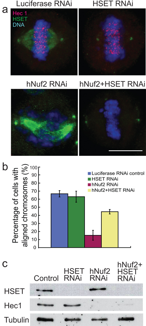
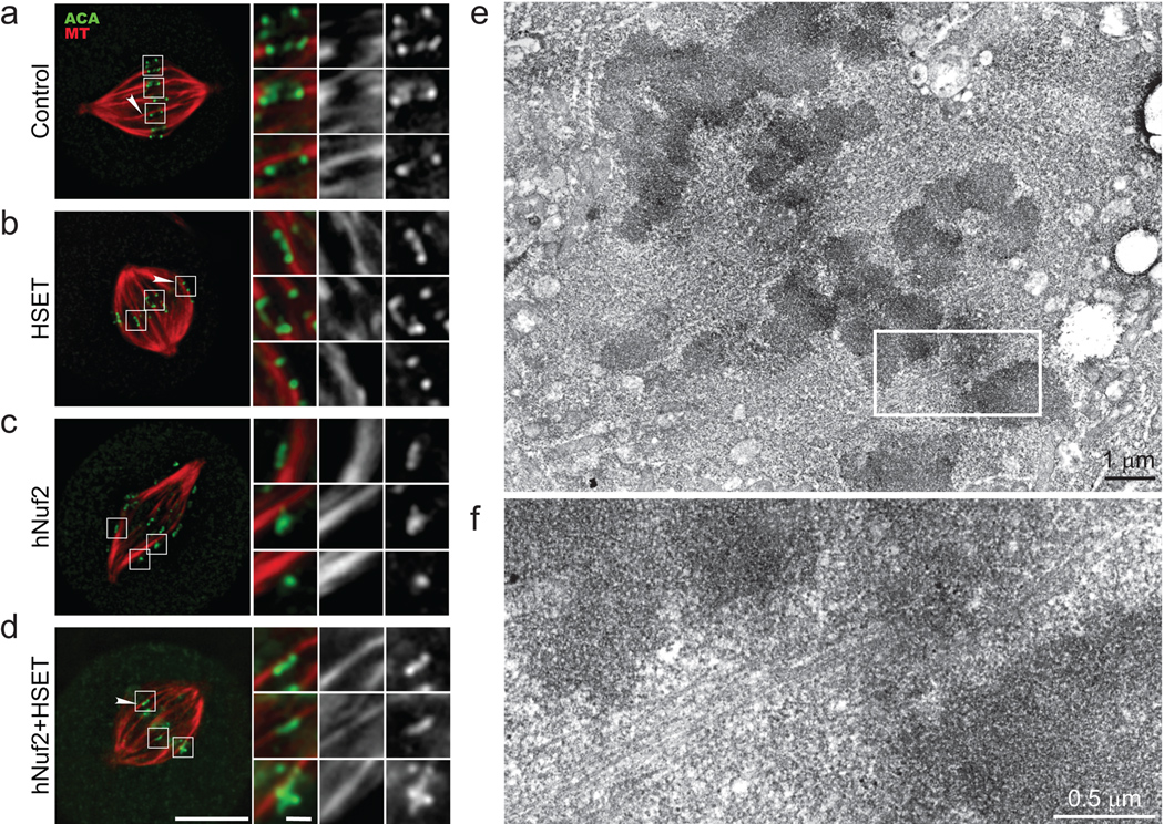
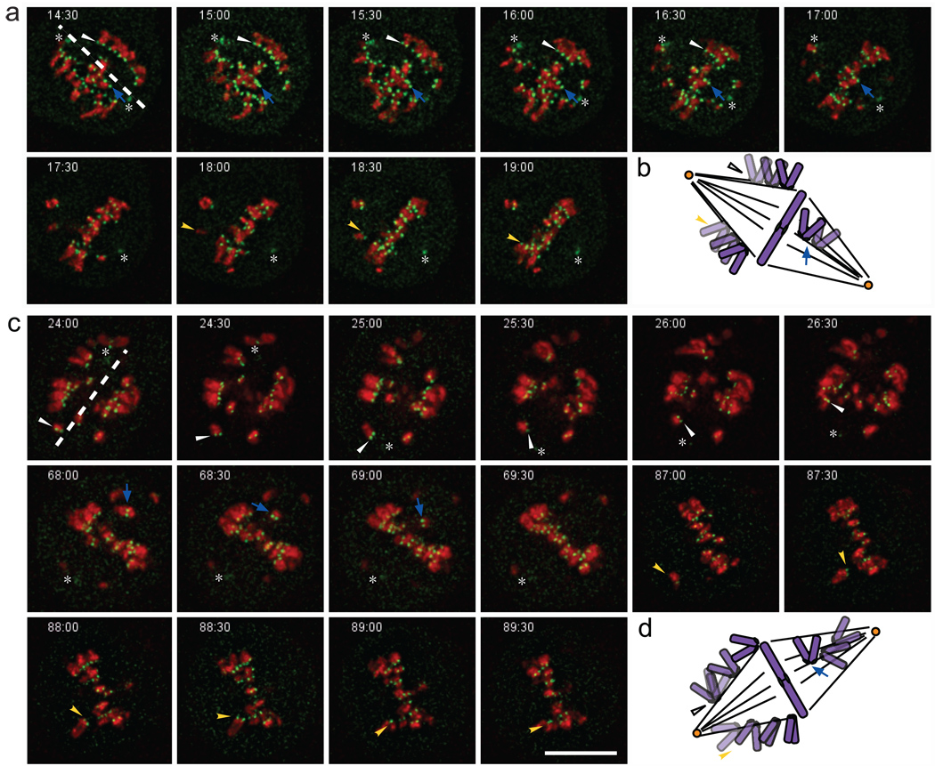
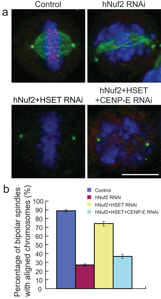
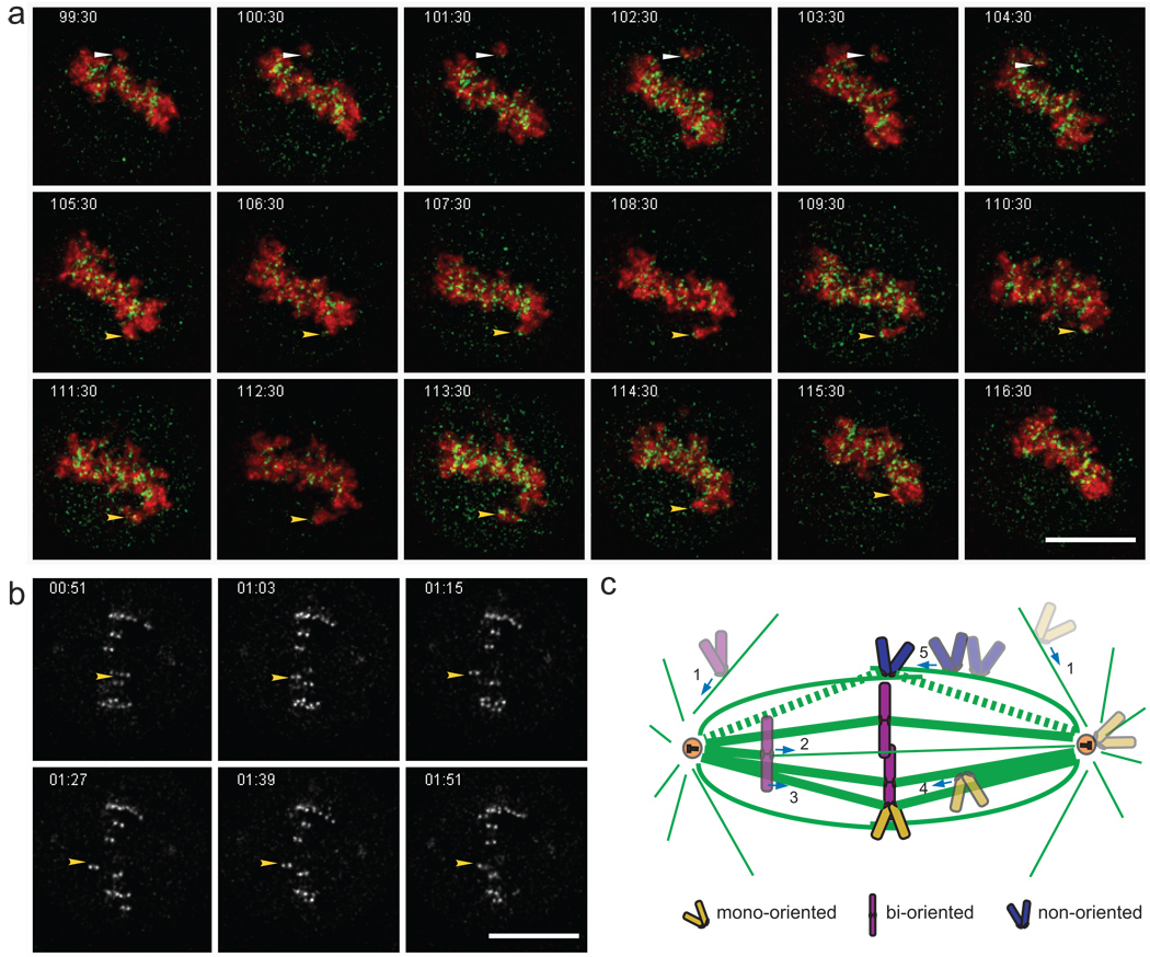
Proper chromosome congression (the process of aligning chromosomes on the spindle) contributes to accurate and faithful chromosome segregation. It is widely accepted that congression requires kinetochore fibres (K-fibres), microtubule bundles that extend from the kinetochores to spindle poles. Here, we demonstrate that chromosomes in human cells co-depleted of HSET (human kinesin-14) and hNuf2 (human Ndc80/Hec1-complex component) can congress to the metaphase plate in the absence of K-fibres. However, the chromosomes are not stably maintained at the metaphase plate under these conditions. Chromosome congression in HSET + hNuf2 co-depleted cells required the plus-end directed motor CENP-E (centromere protein E; kinesin-7 family member), which has been implicated in the gliding of mono-oriented kinetochores alongside adjacent K-fibres. Thus, proper end-on attachment of kinetochores to microtubules is not necessary for chromosome congression. Instead, our data support the idea that congression allows unattached chromosomes to move to the middle of the spindle where they have a higher probability of establishing connections with both spindle poles. These bi-oriented connections are also used to maintain stable chromosome alignment at the spindle equator.


Figures

Figure 1
Figure 1. Chromosomes can align in cells lacking K-fibres
(a) HeLa cells transfected with luciferase control RNAi oligo (Control), hNuf2 RNAi oligo, or hNuf2+HSET RNAi oligo and stained to visualise HSET (green) Hec1 (red), and DNA (blue). Scale bar, 10 µm. (b) The average percentage of prometaphase and metaphase cells with aligned chromosomes is graphed ± s.d. from three independent experiments where ~100 mitotic cells were scored per experiment. The differences between all the pairs are significant (p<0.05) (c) Western blot analysis of samples from each knockdown probed with antibodies to HSET, Hec1, and tubulin.
Figure 2
Figure 2. Kinetochores laterally bind stabilised MT bundles in the absence of K-fibres
HeLa cells in control (a), HSET (b), hNuf2 (c), or hNuf2+HSET (d) RNAi were treated with 0.2 mM CaCl2 to depolymerise highly dynamic MTs and stained to visualise MTs (red), kinetochores (ACA, green) and DNA (blue). Scale bar, 10 µm for whole spindle images (left) or 1 µm for insets (right). Arrowheads indicate a MT bundle that transverses the spindle equator without making an end-on attachment to a kinetochore. (e) Correlative light/serial section EM was performed in hNuf2+HSET RNAi cells. Low magnification view of a cell showing the spindle equator region. (f) Higher magnification view of the region boxed in panel (e). Notice a bundle of microtubules bypassing kinetochores.
Figure 3
Figure 3. In the absence of K-fibres, kinetochores congress with alternating kinetochores leading
Triple labelled HeLa cells (GFP-CENP-A, GFP-gamma-tubulin, and mcherry-H2B) treated with control luciferase (a) or hNuf2+HSET (c) RNAi were imaged at 30 s interval from nuclear envelope breakdown until telophase and analysed for kinetochore orientation during congression. The time stamp is marked in the upper left of each panel, asterisks mark the position of spindle poles, and the coloured arrowheads mark individual kinetochore pairs schematised in (b) and (d). Scale bar, 10 µm. Note that the arrow heads that reflect the orientation of the kinetochore pairs in the control cells do not change much over time, whereas the arrow heads in the hNuf2+HSET RNAi cell change orientation dramatically with time.
Figure 4
Figure 4. Chromosome congression in cells with disrupted K-fibres is dependent on CENP-E
(a) Cells in which hNuf2, HSET, and CENP-E were knocked down were stained to visualize HSET (green), Hec1 (red) and DNA (blue). Scale bar, 10 µm. (b) Quantification of the percentage of bipolar spindles with aligned chromosomes from the four different RNAi conditions. The mean percentages of spindles ± s.d. were graphed for each condition. Three independent experiments and approximately 300 total cells were scored per RNAi condition. The differences between the mean percentages of control versus hNuf2 RNAi, hNuf2 RNAi versus hNuf2+HSET RNAi and hNuf2+HSET versus hNuf2+HSET+CENP-E RNAi are all significant (p<0.05). The difference between hNuf2 RNAi and hNuf2+HSET+CENP-E RNAi is not significant (p=0.45).
Figure 5
Figure 5. K-fibres are needed to maintain chromosome alignment
(a) Triple labelled HeLa cells were analysed for chromosome movement at 30 s intervals after congression in hNuf2+HSET double RNAi cells. The timestamp is shown in the upper left panel of each panel, and white and yellow arrowheads point to two distinct unstable chromosomes that move off the metaphase plate and then congress back to the metaphase plate. (b) Kinetochore movements were tracked in cells with disrupted K-fibres at 3 s intervals during congression in hNuf2+HSET RNAi cells. The yellow arrowhead corresponds to a chromosome that displays a rapid movement away from and back toward the metaphase plate. (c) Model diagramming the different pathways by which chromosomes congress to the metaphase plate. Chromosome 1 (purple) exhibits rapid lateral movement toward the spindle pole and then becomes bi-oriented and congresses through activities at its kinetochore (2) or its chromosome arms (3). Alternatively, chromosome 1 (light yellow) can congress via the mono-oriented pathway (4). Our data suggest that chromosomes (blue) do not need to be specifically oriented but are still able to congress on any stabilised MT bundle (5).

Comment in

References

    1. Maiato H, DeLuca J, Salmon ED, Earnshaw WC. The dynamic kinetochore-microtubule interface. J. Cell Sci. 2004;117:5461–5477. - PubMed
    1. Walczak CE, Heald R. Mechanisms of mitotic spindle assembly and function. Int. Re.v Cytol. 2008;265:111–158. - PubMed
    1. Cai S, Weaver LN, Ems-McClung SC, Walczak CE. Kinesin-14 family proteins HSET/XCTK2 control spindle length by cross-linking and sliding microtubules. Mol. Biol. Cell. 2009;20:1348–1359. - PMC - PubMed
    1. Manning AL, Compton DA. Mechanisms of spindle-pole organization are influenced by kinetochore activity in mammalian cells. Curr. Biol. 2007;17:260–265. - PubMed
    1. DeLuca JG, Moree B, Hickey JM, Kilmartin JV, Salmon ED. hNuf2 inhibition blocks stable kinetochore-microtubule attachment and induces mitotic cell death in HeLa cells. J. Cell Biol. 2002;159:549–555. - PMC - PubMed

Publication types

MeSH terms