. 2009 Jul;12(7):864-71.
doi: 10.1038/nn.2346. Epub 2009 Jun 14.

Pathogenic huntingtin inhibits fast axonal transport by activating JNK3 and phosphorylating kinesin

Affiliations

Pathogenic huntingtin inhibits fast axonal transport by activating JNK3 and phosphorylating kinesin

Gerardo A Morfini et al. Nat Neurosci. 2009 Jul.

Abstract

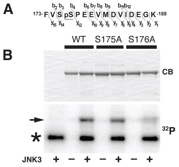
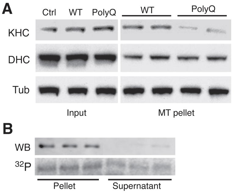
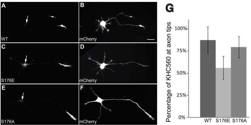
Selected vulnerability of neurons in Huntington's disease suggests that alterations occur in a cellular process that is particularly critical for neuronal function. Supporting this idea, pathogenic Htt (polyQ-Htt) inhibits fast axonal transport (FAT) in various cellular and animal models of Huntington's disease (mouse and squid), but the molecular basis of this effect remains unknown. We found that polyQ-Htt inhibited FAT through a mechanism involving activation of axonal cJun N-terminal kinase (JNK). Accordingly, we observed increased activation of JNK in vivo in cellular and mouse models of Huntington's disease. Additional experiments indicated that the effects of polyQ-Htt on FAT were mediated by neuron-specific JNK3 and not by ubiquitously expressed JNK1, providing a molecular basis for neuron-specific pathology in Huntington's disease. Mass spectrometry identified a residue in the kinesin-1 motor domain that was phosphorylated by JNK3 and this modification reduced kinesin-1 binding to microtubules. These data identify JNK3 as a critical mediator of polyQ-Htt toxicity and provide a molecular basis for polyQ-Htt-induced inhibition of FAT.


Figures

Figure 1
Figure 1. Endogenous Htt does not interact with molecular motors
A) Detergent-soluble brain lysates obtained from wild type (WT) and 14 month-old HdhQ109 knock-in (polyQ) mice were immunoprecipitated with antibodies against Htt, kinesin-1 (KHC), or dynein intermediate chains (DIC). Immunoblots of resulting immunoprecipates (IPP) showed that anti-kinesin-1 antibodies (H213) effectively precipitated both kinesin-1 heavy (KHC) and light chains (KLC), but failed to precipitate Htt, dynein heavy chain (DHC) or dynein intermediate chain (DIC). Similarly, anti-DIC antibodies (74.116) immunoprecipitated DIC and DHC, but not Htt. Conversely, anti-Htt antibodies effectively immunoprecipitated Htt, but not KHC, KLC, DIC or DHC. Immunoprecipitation with non-immune mouse IgG (NMIgG) provided a control for non-specific immunoprecipitation. An aliquot of each brain lysate before immunoprecipitation (Input) was used as a positive control. B) Detergent-soluble brain lysates from wild type (WT-Htt) and HdhQ109 knock-in (polyQ-Htt) mice were subjected to three cycles of immunoprecipitation with antibodies against kinesin-1. Aliquots of each supernatant (SN1-3) were analyzed by immunoblot. Note reductions in both KHC and KLC immunoreactivity with each cycle. In contrast, no change in Htt levels was detected, regardless of mouse genotype. Immunoprecipitations with a non-immune mouse IgG (Ctrl) served as a control for non-specific precipitation of proteins.
Figure 2
Figure 2. JNK inhibitors prevent polyQ-Htt-induced FAT inhibition
Vesicle motility assays in isolated axoplasm show individual velocity measurements (arrowheads) as a function of time. Dark arrowheads and line show anterograde kinesin-dependent FAT rates and reverse grey arrowheads and line show retrograde, cytoplasmic dynein-dependent FAT rates. In vitro translated Htt constructs were perfused as before. A) Perfusion of WT-Htt does not affect FAT B) Perfusion of polyQ-Htt inhibited both anterograde and retrograde FAT. C) Endogenous JNK activity in squid axoplasm is revealed by in vitro phosphorylation assays. A time-dependent increase in cJun phosphorylation at JNK sites (cJun S63/73) is seen when GST-cJun is incubated with squid axoplasm lysates (Axo). Phosphorylation of GST-cJun is completely inhibited by either SP600125 or JIP peptide (JIP), two well-characterized JNK inhibitors. Anti-GST antibody shows similar GST-cJun levels for each reaction (cJun Total). Co-perfusion of polyQ-Htt with either D) SP600125 (500nM) or E) JIP peptide (100 μM) completely prevented inhibition of FAT elicited by polyQ-Htt (compare D-E with B). E) In contrast, co-perfusion of polyQ-Htt with the histone deacetylase inhibitor BC-6-25 (10μM) failed to prevent polyQ-Htt effects on FAT. These results indicate that polyQ-Htt-mediated inhibition of FAT involves activation of endogenous axoplasmic JNK.
Figure 3
Figure 3. PolyQ-Htt increases JNK activity in Huntington’s disease models
JNK activation was evaluated by immunoblots with active JNK (pJNK) and total JNK (pan-JNK) antibodies, which mainly recognized bands at p54 and p46. A) NSC34 cells transfected with WT-Htt (18Q) or polyQ-Htt (56Q) constructs were analyzed after 24h. Both endogenous (End) and exogenous (Exo) Htt were detectable. Total JNK (Pan-JNK) was similar for untransfected (Ctrl), WT-Htt (WT) and polyQ-Htt (polyQ) transfected cells, but phosphorylated p54 JNK (pJNK) increased with polyQ-Htt. B) JNK1, JNK2 and JNK3 antibody specificity were validated with GST-JNKs (left), and striatal lysates with individual JNK genes ablated (right). JNK1, JNK2 and JNK3 antibodies showed no immunoreactivity in brains from corresponding knockout samples. C) Striata from wild type (WT), heterozygous (Hetero) and homozygous (Homo) HdhQ109 mice showed similar Total JNK (pan-JNK) regardless of genotype, whereas active JNK (pJNK) increased in HdhQ109 mice. Akt activation (p-Akt) was similar for all mice. D) Immunoblots using NSC34 (top) and mouse brain (bottom) showed JNK1 co-migrating with p46, whereas JNK2 and JNK3 co-migrated with p54. E) Quantitative analysis of immunoblots in C showed activation of both p54 and p46 JNK in HdhQ109 mice. pJNK/JNK ratios for JNK1 (p46) or JNK2/3 (p54) indicated that both JNK2/3 and JNK1 activity were higher in homozygous than heterozygous HdhQ109 mice. JNK2/3 was activated to a greater extent than JNK1, suggesting differential activation of JNKs by polyQ-Htt. Differences between JNK2/3 activity in WT and HdhQ109 were significant (p ≤ 0.01 in a pooled t-test).
Figure 4
Figure 4. The effects of polyQ-Htt on FAT are mediated by JNK3
SB203580 blocks the action of pathogenic androgen receptor on FAT, but SB203580 is a poor inhibitor of JNK1 (IC50≥100 μM) and a good inhibitor of JNK2/3 (IC50≈10μM). A)In vitro phosphorylation assays in axoplasm showed a dose-dependent inhibition of axoplasmic JNK activity by SB203580. Notably, significant JNK activity remained in the presence of 10μM SB203580, presumably representing endogenous JNK1 activity. B) In contrast, vesicle motility assays in isolated squid axoplasm show that effects of polyQ-Htt on FAT were blocked by SB203580 at 10μM, which is consistent with an action on JNK2/3. C) Sequence alignment of a JIP peptide inhibitor (Calbiochem) with portions of JSAP1a and JSAP1d polypeptides. The boxed region indicates the highly conserved JNK binding domain (JBD). A 31-aminoacid peptide sequence unique to JSAP1d (underlined) selectively interferes with binding to JNK3. D) Co-perfusion of polyQ-Htt with JSAP1a (5 μM) which blocks both JNK1 and JNK3 activity, effectively prevents effects of polyQ-Htt on FAT. E) Co-perfusion of polyQ-Htt with JSAP1d (5μM), which preferentially inhibits JNK1, only partially blocks polyQ-Htt effects. These results suggest that JNK3 mediates the effects of polyQ-Htt on FAT.
Figure 5
Figure 5. Active JNK3 mimics the effect of polyQ-Htt on FAT
Effects of active, recombinant JNK1, JNK2 and JNK3 were evaluated by vesicle motility assays in isolated axoplasm. A) Perfusion of active JNK1 kinase (200nM) in axoplasm had no effect on FAT. B) Perfusion of active JNK2 (100nM) slightly inhibited anterograde FAT, but had no significant effect on retrograde FAT. C) Perfusion of JNK3 (100nM) significantly inhibited FAT in both directions, as observed with polyQ-Htt (compare to Fig. 2B) D) Bar graphs showing mean FAT rates in axoplasm perfused with active JNKs and polyQ-Htt. Error bars show SEM. Data represent pooled FAT rate measurements between 30-50 min of observation. Anterograde FAT rates after polyQ-Htt perfusion were comparable to those elicited by perfusion of active JNK3 and both JNK3 and polyQ-Htt significantly inhibit retrograde FAT.
Figure 6
Figure 6. JNK3 phosphorylates kinesin-1 heavy chains at Ser176
A) Mass spectrometry studies identified a tryptic peptide within the motor domain of kinesin-1 (amino acids 173-188) showing unequivocal evidence of phosphorylation by JNK3 (see also Supplemental Fig. 2). B) Recombinant KHC584-WT protein was incubated in the presence (+) or absence (-) of active JNK3 and 32P-ATP, and analyzed by SDS-PAGE. The ability of JNK3 to phosphorylate KHC584 at S175 and S176 was also evaluated using KHC584-S175 and KHC584-S176A mutants. An autoradiogram (32P) showed a dramatic reduction in the phosphorylation of a KHC584-S176A (S176A) construct relative to wild type (WT) and KHC584-S175A (S175A) constructs (arrow). Coomassie blue (CB) staining showed comparable amounts of KHC584 in all lanes. Asterisk indicates autophosphorylated JNK3. These results indicated that S176 is the main residue phosphorylated by JNK3.
Figure 7
Figure 7. PolyQ-Htt expression inhibits kinesin-1 binding to microtubules
A) Lysates of NSC34 cells transfected with WT-Htt or polyQ-Htt as in Fig. 3A were analyzed by immunoblot. Total levels (Input) of kinesin-1 (KHC), dynein heavy chain (DHC) and tubulin (Tub) were comparable for untransfected (Ctrl), WT-Htt and polyQ-Htt-expressing cells. However, the fraction of kinesin-1 recovered in association with microtubules was reduced for lysates from polyQ-Htt expressing cells, when compared to untransfected and WT-Htt-expressing cells. B) Microtubule-binding assays using recombinant kinesin-1 (KHC584). Immunoblot (WB) shows that unphosphorylated KHC584 is mainly recovered in association with microtubules (Pellet). An autoradiogram (32P) reveals a significant fraction of JNK3-phosphorylated KHC584 remains in the supernatant.
Figure 8
Figure 8. A mutation mimicking Ser176 phosphorylation reduces the translocation efficiency of constitutively active kinesin-1 in cultured hippocampal neurons
A-F: Representative stage 3 neurons were co-transfected with GFP-tagged constructs encompassing the first 560 amino acids of kinesin-1 (KHC560, A, C, and E) and mCherry, a soluble marker protein that diffuses throughout the cell (B, D, and F, scale bar= 20μm). Cells were fixed with formaldehyde and imaged 5 hours after transfection. Wild type (WT, A) and non-phosphorylatable S176A mutant (S176A, E) constructs efficiently translocated so that they were localized almost exclusively at the axon tip. In contrast, a significant fraction of the pseudophosphorylated S176 mutant (S176E, C) was found throughout the cell body. Arrowheads in A, C and E indicate the location of the neuronal cell bodies as seen in B, D, and F. G: The percentage of total KHC560-GFP fluorescence localized at the axon tip was measured in cells expressing KHC560 WT (N=22), KHC560 S176E (N=33) or KHC560 S176A (N=30). Significantly less KHC560 S176E accumulated at axon tips with the wild-type construct (t-test, p<0.0001); only marginally less KHC560 S176A accumulated at the axon tip than the wild-type construct (t-test, p~0.05). Bars show mean and standard deviation.

References

    1. Okun MS. Huntington’s disease: what we learned from the original essay. Neurologist. 2003;9:175–179. - PubMed
    1. Orr HT, Zoghbi HY. Trinucleotide repeat disorders. Annu Rev Neurosci. 2007;30:575–621. - PubMed
    1. Morfini G, Pigino G, Brady ST. Polyglutamine Expansion Diseases: Failing to Deliver. Trends Molec Med. 2005;11:64–70. - PubMed
    1. Gunawardena S, et al. Disruption of axonal transport by loss of huntingtin or expression of pathogenic polyQ proteins in Drosophila. Neuron. 2003;40:25–40. - PubMed
    1. Lee WC, Yoshihara M, Littleton JT. Cytoplasmic aggregates trap polyglutamine-containing proteins and block axonal transport in a Drosophila model of Huntington’s disease. Proc Natl Acad Sci U S A. 2004;101:3224–3229. - PMC - PubMed

Publication types

MeSH terms