

Pontine infarction with pure motor hemiparesis or hemiplegia: a prospective study
- PMID: 19527495
- PMCID: PMC2707361
- DOI: 10.1186/1471-2377-9-25
Pontine infarction with pure motor hemiparesis or hemiplegia: a prospective study
Abstract
Background: The study aimed to prospectively observe the clinical and neuroimaging features of pontine infarction with pure motor hemiparesis (PMH) or hemiplegia at early stage.
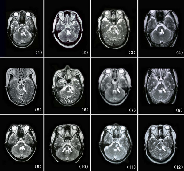
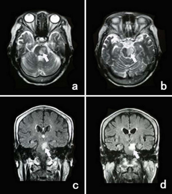
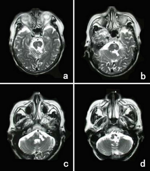
Methods: In 118 consecutive selected patients with the first-ever ischemic stroke within 6 hours after onset, fifty of them presented with PMH or hemiplegia and had negative acute computed tomography (CT) scans, then magnetic resonance imaging (MRI) confirmed the corresponding infarcts in pons or cerebrum. The clinical and neuroimaging features of the pontine infarctions were compared with those of cerebral infarctions.
Results: The pontine infarction with PMH or hemiplegia accounted for 10.2% (12/118) of all first-ever ischemic stroke patients and 24% (12/50) of the patients with both PMH or hemiplegia and acute negative CT scans. Compared to the patients with cerebral infarction, the patients with pontine infarction had more frequency of diabetes mellitus (50.0% vs 5.3%, P = 0.001), nonvertiginous dizziness at onset (58.3% vs 21.1%, P = 0.036) and a progressive course (33.3% vs 2.6%, P = 0.011).
Conclusion: The pontine infarction may present as PMH or hemiplegia with more frequency of nonvertiginous dizziness, a progressive course and diabetes mellitus. MRI can confirm the infarct location in the basal pons at early stage after stroke onset.
Figures
References
-
- Silverman IE, Liu GT, Volpe NJ, Galetta SL. The crossed paralyses. The original brain-stem syndromes of Millard-Gubler, Foville, Weber, and Raymond-Cestan. Arch Neurol. 1995;52:635–638. - PubMed
-
- Bassetti C, Bogousslavsky J, Barth A, Regli F. Isolated infarcts of the pons. Neurology. 1996;46:165–175. - PubMed
-
- Fisher CM, Curry HB. Pure motor hemiplegia of vascular origin. Arch Neurol. 1965;13:30–44. - PubMed
-
- Heo JH, Bang OY, Choi SA. Pure motor hemiplegia with conjugate lateral gaze palsy in pontine lacunar infarction. Yonsei Med J. 1996;37:86–88. - PubMed
-
- Kataoka S, Hori A, Shirakawa T, Hirose G. Paramedian pontine infarction Neurological/topographical correlation. Stroke. 1997;28:809–815. - PubMed
Publication types
MeSH terms
LinkOut - more resources
Full Text Sources
