Review
. 2009 Jun 18;5(6):527-49.
doi: 10.1016/j.chom.2009.05.016.

Autophagy, immunity, and microbial adaptations

Affiliations
Review

Autophagy, immunity, and microbial adaptations

Vojo Deretic et al. Cell Host Microbe. .

Abstract

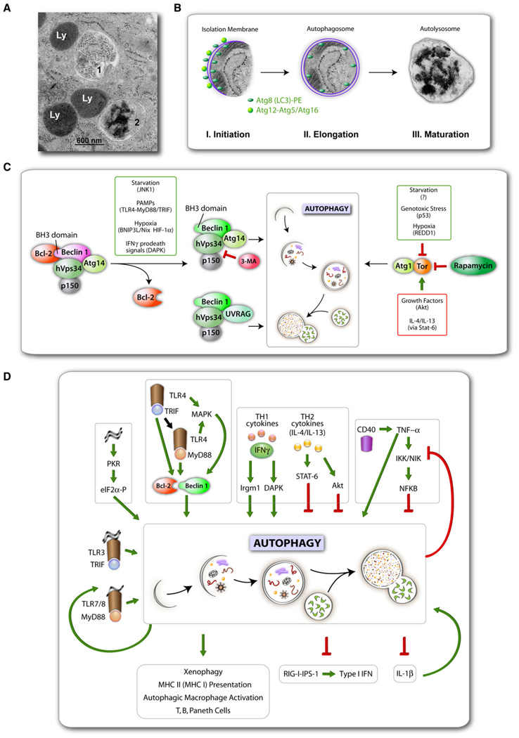
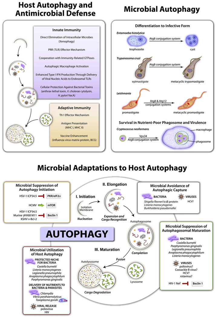
Autophagy adjusts cellular biomass and function in response to diverse stimuli, including infection. Autophagy plays specific roles in shaping immune system development, fueling host innate and adaptive immune responses, and directly controlling intracellular microbes as a cell-autonomous innate defense. As an evolutionary counterpoint, intracellular pathogens have evolved to block autophagic microbicidal defense and subvert host autophagic responses for their survival or growth. The ability of eukaryotic pathogens to deploy their own autophagic machinery may also contribute to microbial pathogenesis. Thus, a complex interplay between autophagy and microbial adaptations against autophagy governs the net outcome of host-microbe encounters.


Figures

Figure 1
Figure 1. Autophagy Stages, Regulation, and Roles in Immunity
(A) Electron micrograph comparing the appearance of an autophagosome (1) versus autolysosome (2) with conventional lysosomes (Ly). (B) A composite of autophagic membrane formation (schematic) with actual autophagosome from an electron micrograph. Three stages can be discerned by ultrastructural morphology: initiation (crescent membrane decorated on both ssides with Atg protein-lipid [Atg8-PE; known as LC3-II] and protein-protein conjugates [Atg12-Atg5, complexed with Atg16]), elongation (growth of isolation membrane) ending in its closure to form an autophagosome, and maturation that involves the formation of degradative autolysosomes through fusion of autophagosomes with lysosomal organelles (electron-dense granular material represents ribosomal degradation intermediates). All micrographs in (A) and (B) are courtesy of Eeva-Lisa Eskalinen (reproduced with permission) (C) Two signaling systems control induction of autophagy: left, hVps34-Beclin 1; right, Tor-Atg1. Various signals and systems transmitting them that lead to autophagy activation are shown in smaller boxes. Rapamycin induces autophagy by inhibiting Tor, and some immune signals that are transmitted via TLR adapters MyD88 and TRIF or DAPK downstream of IFNγ lead to activation of Beclin 1 (complexed with the phosphatidylinositol 3 kinase hVps34) by its dissociation from Bcl-2. Note that starvation, a classical inducer of autophagy, affects both Bcl-2-Beclin 1 complex (via JNK1) and Tor activity. Th2 cytokines and many growth factors in general inhibit autophagy via Tor (or in the case of immunologically induced autophagy, e.g., by IFNγ, via Stat-6 downstream of IL-4/IL-13). (D)Immunological inputsand outputs of autophagy. Xenophagy,autophagic Macrophage activation, additional terms, factors, and relationships are described in the text.
Figure 2
Figure 2. Autophagy in Inflammation
Top shows two autophagy factors identified as Crohn’s disease (a form of inflammatory bowel disease) susceptibility loci. The IRGM gene has single nucleotide polymorphisms (SNP)and a 20 kb deletion in the promoter region associated with CD. ATG16L1 alleles encode either a protective ATG16L1*300T or a risk form of ATG16L1*300A (CCD, coiled coil domain; green boxes, WD repeats domain [absent in yeast Atg16]). Middle panel shows intestinal epithelium with different cell types along with the proposed functions of autophagy in the ileal epithelium. Side box shows that thymic selection of naive T cell repertoires depends on autophagy, and autophagic anomalies may contribute to inflammation at peripheral sites such as intestinal mucosa. Bottom box lists 3 different effects reported for Atg16L1 (mouse) or ATG16L1 (human epithelial cells) mutations. ATG16L1*300A has been tested in cell lines. Atg16L1HM, hypomorphic Atg16L1 allele, and Atg16LΔCCD construct have been tested in vivo in transgenic mice.
Figure 3
Figure 3. Schematic Depicting Functions of Host Autophagy in Antimicrobial Defense, Functions of Microbial Autophagy, and Microbial Adaptations to Host Autophagy
See Table 1 and Table 2 for further details of specific host autophagy-pathogen interactions.

References

    1. Ait-Goughoulte M, Kanda T, Meyer K, Ryerse JS, Ray RB, Ray R. Hepatitis C virus genotype 1a growth and induction of autophagy. J. Virol. 2008;82:2241–2249. - PMC - PubMed
    1. Al-Younes HM, Brinkmann V, Meyer TF. Interaction of Chlamydia trachomatis serovar L2 with the host autophagic pathway. Infect. Immun. 2004;72:4751–4762. - PMC - PubMed
    1. Al-Zeer MA, Al-Younes HM, Braun PR, Zerrahn J, Meyer TF. IFN-gamma-inducible Irga6 mediates host resistance against Chlamydia trachomatis via autophagy. PLoS ONE. 2009;4:e4588. - PMC - PubMed
    1. Alonso S, Pethe K, Russell DG, Purdy GE. Lysosomal killing of Mycobacterium mediated by ubiquitin-derived peptides is enhanced by autophagy. Proc. Natl. Acad. Sci. USA. 2007;104:6031–6036. - PMC - PubMed
    1. Alvarez VE, Kosec G, Sant’Anna C, Turk V, Cazzulo JJ, Turk B. Autophagy is involved in nutritional stress response and differentiation in Trypanosoma cruzi. J. Biol. Chem. 2008;283:3454–3464. - PubMed

Publication types