

Review
doi: 10.1098/rstb.2008.0312.
See it with feeling: affective predictions during object perception
Affiliations
- PMID: 19528014
- PMCID: PMC2666711
- DOI: 10.1098/rstb.2008.0312
Item in Clipboard
Review
See it with feeling: affective predictions during object perception
Philos Trans R Soc Lond B Biol Sci.
.
Abstract
People see with feeling. We 'gaze', 'behold', 'stare', 'gape' and 'glare'. In this paper, we develop the hypothesis that the brain's ability to see in the present incorporates a representation of the affective impact of those visual sensations in the past. This representation makes up part of the brain's prediction of what the visual sensations stand for in the present, including how to act on them in the near future. The affective prediction hypothesis implies that responses signalling an object's salience, relevance or value do not occur as a separate step after the object is identified. Instead, affective responses support vision from the very moment that visual stimulation begins.
Figures
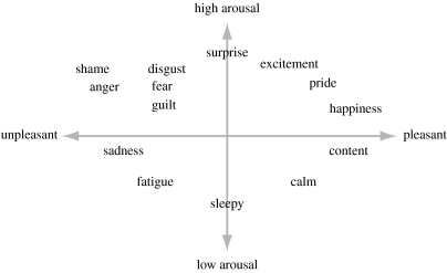
A descriptive map of core affect.
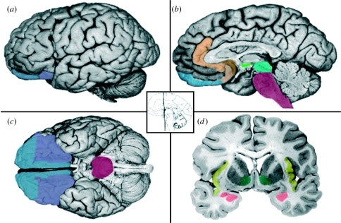
(a–d) The affective workspace. The neural workspace for core affect includes a broadly distributed set of interconnected cortical and subcortical brain areas that are not functionally specific to affect, but that realize affective responses as a network (for imaging evidence, see Kringelbach & Rolls 2004; Barrett et al. 2007a; Wager et al. 2008; for a discussion, see Barrett & Bliss-Moreau in press). Some areas are traditionally considered to be ‘emotional’, such as the amygdala (rose) and ventral striatum (green). Other areas were (until recently) considered ‘cognitive’ (cf. Duncan & Barrett 2007; for a similar view, see Pessoa 2008), such as the orbitofrontal cortex (OFC; blue and purple). Typically, researchers use the term ‘orbital frontal cortex’ to refer to anywhere within the orbital sector of the prefrontal cortex. This includes the lateral parts (blue) that are bounded by the ventrolateral prefrontal cortex (vlPFC) and agranular insula (yellow), as well as the medial portions that wrap to include the ventromedial prefrontal cortex (vmPFC; purple) extending back to the sub/pregenual portions of the anterior cingulate cortex (ACC; brown and gold). Other components of this workspace include the hypothalamus (light green) and autonomic control centres in the midbrain and brainstem (turquoise and maroon). This neural reference space for effect is meant to be non-specific without sounding vague, in that different assemblies of neurons across these areas realize momentary representations of value (Ghashghaei & Barbas 2002; Barbas et al. 2003). Photographs taken from DeArmond et al. (1989), pp. 5, 7, 8 and 43.
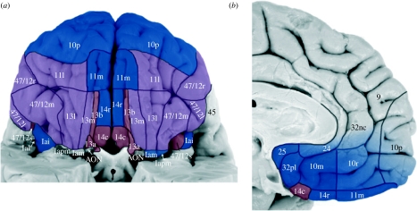
(a) Medial and (b) lateral networks of the OFC. Adapted from Öngür et al. (2003) based on the evidence from Barbas (1993, 2000), Carmichael & Price (1995), McDonald (1998), Cavada et al. (2000), Ghashghaei & Barbas (2002) and Stefanacci & Amaral (2002). Orbital and medial views of the brain are shown. The medial network is shown in blue and has robust reciprocal connections to all limbic areas (including many nuclei within the amygdala and the ventral striatum), as well as to the hypothalamus, midbrain, brainstem and spinal cord areas that are involved in internal state regulation. The medial OFC has few direct connections to sensory cortices. The lateral network is shown in purple and has robust connections with unimodal sensory association areas as well as the cortical aspects of the amygdala (including the basolateral complex, which also receives sensory input from unimodal association areas). The lateral OFC has few direct projections to autonomic and endocrine control centres in the hypothalamus, midbrain and brainstem, but has some influence on those autonomic centres via projections to the intercalated masses within the amygdala that have the net effect disinhibiting (i.e. activating) these nuclei. The areas that connect the two networks are shown in rose.
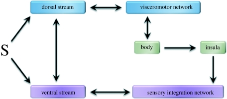
Connections between the OFC and visual networks. ‘S’ represents visual information that reaches the dorsal ‘where’ stream and the ventral ‘what’ stream both from early visual areas and from the thalamus. The medical OFC is part of a ‘visceromotor’ network that helps to control internal body state. The lateral OFC is part of a ‘sensory integration network’ that joins together sensory representations from outside and inside the body.
References
-
- Abramson B.P., Chalupa L.M. The laminar distribution of cortical connections with the tecto- and cortico-recipient zones in the cat's lateral posterior nucleus. Neuroscience. 1985;15:81–95. doi:10.1016/0306-4522(85)90125-3 - DOI - PubMed
-
- Arnold M.B. Columbia University Press; New York, NY: 1960. Emotion and personality.
-
- Bar M. A cortical mechanism for triggering top-down facilitation in visual object recognition. J. Cogn. Neurosci. 2003;15:600–609. doi:10.1162/089892903321662976 - DOI - PubMed
-
- Bar M. Visual objects in context. Nat. Rev. Neurosci. 2004;5:617–629. doi:10.1038/nrn1476 - DOI - PubMed
-
- Bar M. The proactive brain: using analogies and associations to generate predictions. Trends Cogn. Sci. 2007;11:280–289. doi:10.1016/j.tics.2007.05.005 - DOI - PubMed
Publication types
MeSH terms
Grants and funding
LinkOut - more resources
Full Text Sources
Miscellaneous