. 2009 Sep;77(9):3886-93.
doi: 10.1128/IAI.01583-08. Epub 2009 Jun 15.

Loa loa Microfilariae evade complement attack in vivo by acquiring regulatory proteins from host plasma

Affiliations

Loa loa Microfilariae evade complement attack in vivo by acquiring regulatory proteins from host plasma

Karita Haapasalo et al. Infect Immun. 2009 Sep.

Abstract

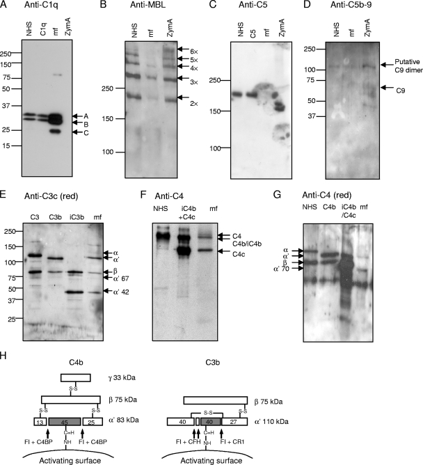
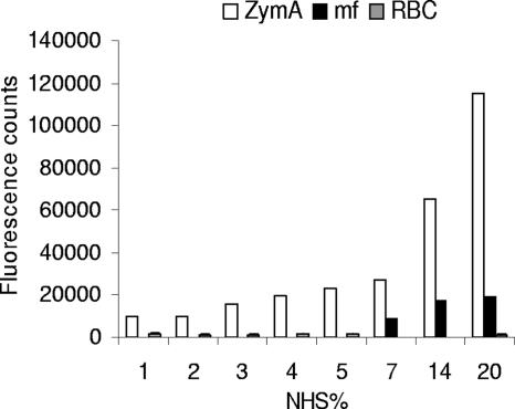
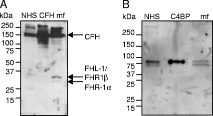
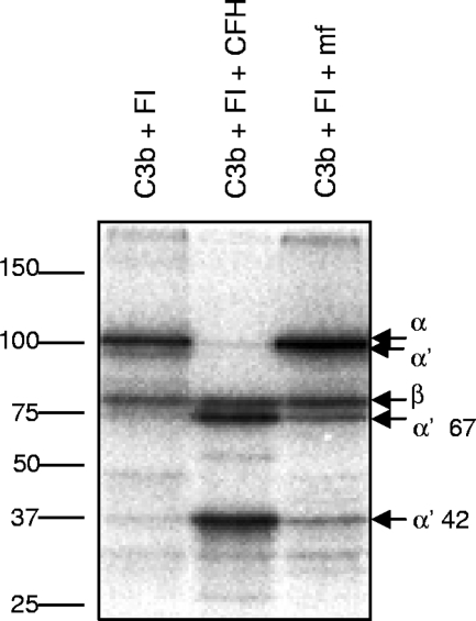
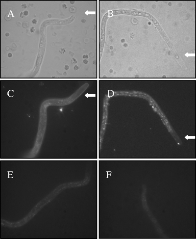
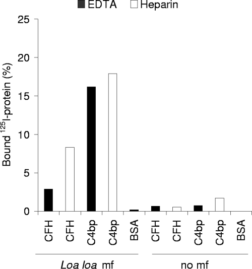
Loa loa is a filarial nematode that infects humans. The adults live in subcutaneous tissues and produce microfilariae that live for several weeks in the blood circulation in order to be transmitted to another person via blood meals of a dipterian vector. As microfilariae live in continuous contact with plasma, it is obvious that they evade the complement system. We studied markers of complement activation and signs of complement regulation on Loa loa microfilariae in vivo. The microfilariae were isolated from anticoagulated blood samples of a Loa loa-infected Caucasian patient. C1q and some mannose-binding lectin but only a limited amount of C3b or C4b fragments and practically no C5 or C5b-9 were present on the microfilariae. The covalently microfilaria-bound C3 and C4 depositions were mainly inactive iC3b, C3c, and iC4b fragments indicating that microfilariae had regulated complement activation in vivo. Also, in vitro deposition of C3b onto the microfilariae upon serum exposure was limited. The patient-isolated microfilariae were found to carry the host complement regulators factor H and C4b-binding protein on the outermost layer, so called sheath. The microfilaria-bound factor H was functionally active. Binding of the complement regulators to the microfilariae was confirmed in vitro using (125)I-labeled factor H and C4b-binding protein. In conclusion, our study shows that Loa loa microfilariae block complement activation and acquire the host complement regulators factor H and C4b-binding protein in blood circulation. This is the first time that binding of complement regulators onto nonviral pathogens has been demonstrated to occur in humans in vivo.


Figures

FIG. 1.
FIG. 1.
Deposition of complement components C1q, MBL, and C5; C5b-9 complexes; and fragments of C3 and C4 on the surface of Loa loa MF. The presence of those molecules on MF was analyzed after subjecting purified patient blood-derived MF to either acidic elution (A, B, and C) or elution with reducing buffer (G) or after homogenizing MF (D, E, and F). The samples obtained were used in SDS-polyacrylamide gel electrophoresis under either reducing (A, D, E, and G) or nonreducing (B, C, and F) conditions and subsequent Western blotting with antibodies against the molecules as indicated above each panel (the anti-C5b-9 panel was blotted with anti-C9 neoepitope antibody). As positive controls, complement components C1q (0.3 μg/ml); C5 (1 μg/ml); C3, C3b, and iC3b (0.02 μg each); C4b and iC4b (0.04 μg each); and NHS were used (diluted 1:500 for panel A, 1:100 for panel B, 1:200 for panel C, and 1:1,000 for panels F and G). NHS was used as a negative control for C5b-9 deposition (D). NHS-exposed zymosan A (ZymA) was used as a positive control in the deposition of the complement components MBL (B) and C5b-9 complex (D) and as a negative control in the deposition of C1q (A). The mobility of molecular size markers is indicated on the left sides. Mobilities of the C1q subunits A, B, and C (A); the oligomers of MBL (B); the activated C5b-9 associated C9 complexes (D); the fragments of C3 (E); and the fragments of C4 (F and G) are indicated. A schematic presentation of C4b and C3b fragments and the cleavage sites of FI is shown in panel H.
FIG. 2.
FIG. 2.
Activation of the AP of complement on Loa loa MF in vitro. Patient blood-isolated Loa loa MF, complement-activating zymosan A yeast particles (ZymA), and complement-nonactivating human red blood cells (RBC) were exposed to increasing concentrations of NHS. The amount of surface-bound C3b and iC3b molecules was measured by staining the cells or MF with rabbit anti-C3c antibody and Alexa 488-conjugated secondary antibody followed by detection of the bound fluorescence using a fluorometer. The fluorescence intensity obtained without any NHS incubation was subtracted as background. The results are shown as means for parallel samples assayed in duplicate.
FIG. 3.
FIG. 3.
Analysis of human complement regulators CFH (A) and C4BP (B) bound on the patient EDTA blood-derived Loa loa MF. The MF homogenate and the positive controls NHS (diluted 1:1,000), 1.5 μg of CFH (A), and 0.02 μg of C4BP (B) were subjected to SDS-polyacrylamide gel electrophoresis using 5 to 15% gradient gels under reducing conditions and used for Western blotting with anti-CFH (A) or anti-C4BP (B) antibodies. The presence of 155-kDa CFH (A) and the 70-kDa α-chain of C4BP (B) can be detected in the MF preparations. The mobility of the molecular size marker is indicated on the left.
FIG. 4.
FIG. 4.
Analysis of cofactor activity of Loa loa MF-bound CFH. The MF were isolated from EDTA-anticoagulated blood by the Percoll gradient method and washed. The cofactor activity of the MF-bound CFH was analyzed by incubating the MF with 125I-labeled C3b and FI (C3b+FI+MF). The positive control sample contained 125I-labeled C3b, FI, and CFH and the negative control only 125I-labeled C3b and FI. The mobility of the molecular weight markers is shown on the left, and the identities of the fragments of C3b are indicated on the right.
FIG. 5.
FIG. 5.
Localization of CFH and C4BP bound on Loa loa MF. The MF were enriched from EDTA-blood by the Percoll gradient method, washed, and subjected to immunofluorescence staining using polyclonal goat anti-CFH (C) or rabbit anti-C4BP (D) antibody and Alexa 488-conjugated secondary antibodies. The photographs were taken from representative MF using bright-field (A and B) or fluorescence (C and D) microscopy. Control photographs with only the secondary antibodies are shown in panels E and F. The typical extended sheath in the anterior end of Loa loa MF is indicated with a arrows in panels A, B, C, and D.
FIG. 6.
FIG. 6.
Loa loa MF bind purified CFH and C4BP. The MF were isolated from EDTA-anticoagulated blood (40 MF/tube) or heparin-anticoagulated blood (100 MF/tube) and incubated with 125I-labeled CFH, C4BP, or BSA for 30 min. The unbound proteins were separated from the bound ones by centrifugation through 20% sucrose. Radioactivity in the pellets and supernatants was measured using a gamma counter. The experiment was performed twice in duplicate, and the mean values are indicated.

References

    1. Ajuh, P. M., J. P. Akue, P. Boutin, S. Everaere, and T. G. Egwang. 1995. Loa loa: structural diversity of a 15-kDa repetitive antigen. Exp. Parasitol. 81145-153. - PubMed
    1. Bain, O., and S. Babayan. 2003. Behaviour of filariae: morphological and anatomical signatures of their life style within the arthropod and vertebrate hosts. Filaria J. 216. - PMC - PubMed
    1. Berggård, K., E. Johnsson, E Morfeldt, J. Persson, M. Stålhammar-Carlemalm, and G. Lindahl. 2001. Binding of human C4BP to the hypervariable region of M protein: a molecular mechanism of phagocytosis resistance in Streptococcus pyogenes. Mol. Microbiol. 42539-551. - PubMed
    1. Chandrashekar, R., U. R. Rao, G. R. Rajasekariah, and D. Subrahmanyam. 1984. Separation of viable microfilariae free of blood cells on Percoll gradients. J. Helminthol. 5869-70. - PubMed
    1. Cooper, N. R. 1975. Enzymatic activity of the second component of complement. Biochemistry 144245-4251. - PubMed

Publication types

Substances