. 2009 Sep;77(9):3696-704.
doi: 10.1128/IAI.00438-09. Epub 2009 Jun 15.

Streptococcus gordonii modulates Candida albicans biofilm formation through intergeneric communication

Affiliations

Streptococcus gordonii modulates Candida albicans biofilm formation through intergeneric communication

Caroline V Bamford et al. Infect Immun. 2009 Sep.

Abstract

The fungus Candida albicans colonizes human oral cavity surfaces in conjunction with a complex microflora. C. albicans SC5314 formed biofilms on saliva-coated surfaces that in early stages of development consisted of approximately 30% hyphal forms. In mixed biofilms with the oral bacterium Streptococcus gordonii DL1, hyphal development by C. albicans was enhanced so that biofilms consisted of approximately 60% hyphal forms. Cell-cell contact between S. gordonii and C. albicans involved Streptococcus cell wall-anchored proteins SspA and SspB (antigen I/II family polypeptides). Repression of C. albicans hyphal filament and biofilm production by the quorum-sensing molecule farnesol was relieved by S. gordonii. The ability of a luxS mutant of S. gordonii deficient in production of autoinducer 2 to induce C. albicans hyphal formation was reduced, and this mutant suppressed farnesol inhibition of hyphal formation less effectively. Coincubation of the two microbial species led to activation of C. albicans mitogen-activated protein kinase Cek1p, inhibition of Mkc1p activation by H(2)O(2), and enhanced activation of Hog1p by farnesol, which were direct effects of streptococci on morphogenetic signaling. These results suggest that interactions between C. albicans and S. gordonii involve physical (adherence) and chemical (diffusible) signals that influence the development of biofilm communities. Thus, bacteria may play a significant role in modulating Candida carriage and infection processes in the oral cavity.


Figures
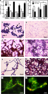
FIG. 1.
FIG. 1.
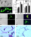
Interactions of C. albicans SC5314 with S. gordonii DL1 in biofilms formed on saliva-coated surfaces. (A) Biomasses of 16 h-biofilms formed at 37°C in 25% human saliva (Saliva), in 25% saliva containing 0.5% glucose (Slv-Glc), in YPT medium, or in YPT medium containing 0.5% glucose (YPT-Glc) by S. gordonii (Sg), C. albicans (Ca), or both S. gordonii and C. albicans (Ca+Sg). The results are representative of three experiments, and the error bars indicate standard deviations. (B) Percentages of cells forming hyphae after 3 h of incubation at 37°C in biofilms for C. albicans grown in 25% saliva containing 0.5% glucose or YPT-Glc, for C. albicans alone (Ca), for C. albicans to which S. gordonii was added (Ca+Sg), and for S. gordonii to which C. albicans was added (Sg+Ca). The results are representative of three experiments, and the error bars indicate standard deviations (*, P = 0.037; **, P = 0.012 [statistically significant]). (C) C. albicans 3-h biofilm in YPT-Glc, showing mixed morphological forms, including blastospores (B), hyphae (H), pseudohyphae (P), and buds (Bu). (D) S. gordonii 3-h biofilm in YPT-Glc, showing chains of cocci with a relatively even surface distribution. (E) Six-hour mixed biofilm in YPT-Glc of antecedent C. albicans (1 h) and S. gordonii, with clusters of streptococci on hyphae (arrows) and around some mother cells. (F) Eight-hour mixed biofilm in YPT-Glc of antecedent S. gordonii (1 h) and C. albicans, showing hyphal networking on a lawn of streptococci (pink). (G) C. albicans 3-h biofilm in 25% saliva containing 0.5% glucose, demonstrating the increased frequency of hyphal formation compared with the biofilm shown in panel C. (H) Eight-hour mixed biofilm in 25% saliva containing 0.5% glucose of antecedent C. albicans (1 h) and S. gordonii, showing high-density hyphal formation and deposition of streptococci. (I) Live/dead staining of C. albicans associated with S. gordonii after 24 h, with metabolically active hyphae (green) containing vacuoles (red). (J) C. albicans and P. aeruginosa PAO1 after 24 h, showing metabolically inactive vesicles (yellow) and dead hyphae.
FIG. 2.
FIG. 2.
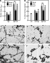
Physical interactions of S. gordonii with C. albicans. (A) Light microscopy (upper panel) and corresponding fluorescence (lower panel) images of fluorescein isothiocyanate-labeled S. gordonii attached to hyphal filaments (H) and mother cells (MC) but not to some blastospores (B) (arrows). (B) Biomasses for 16-h biofilms (formed at 37°C in 25% saliva containing 0.5% glucose [Slv-Glc] or YPT-Glc) of S. gordonii DL1 (Sg), S. gordonii UB1360 Δ(sspA sspB) (Sg sspAB), or antecedent S. gordonii and C. albicans (Sg+Ca). The results are representative of three experiments, and the error bars indicate standard deviations (*, P = 0.0015 [statistically significant]). (C to F) Light microscopy (left panels) and corresponding fluorescence (right panels) images of C. albicans hypha-forming cells mixed with fluorescein isothiocyanate-labeled S. gordonii DL1 (C), S. gordonii UB1360 (D), S. gordonii UB1360/pUB1000sspB+ (E), and S. gordonii UB1545 Δhsa (F). (G and H) Three-hour biofilms of antecedent C. albicans with S. gordonii DL1 (G) or UB1360 (H). The arrow in panel G indicates a close association of S. gordonii DL1 cells with hyphae, which is less apparent in panel H, which is a lower-magnification image showing streptococcal cells distributed across the field.
FIG. 3.
FIG. 3.
Effect of luxS on mixed-species biofilm formation. (A) Biomasses for 16-h monospecies biofilms (37°C in YPT-Glc) of S. gordonii DL1 (Sg wt), the ΔluxS mutant (luxS), or the ΔluxS(luxS+) complemented strain (luxS+) or for S. gordonii DL1 coinoculated with C. albicans (Sg+Ca). The results are representative of three experiments, and the error bars indicate standard deviations (*, P = 0.031 [statistically significant]). (B) Percentages of C. albicans cells forming hyphae (3-h biofilm) alone (Ca), with antecedent C. albicans and S. gordonii (Ca+Sg), or with antecedent S. gordonii and C. albicans (Sg+Ca). The results are representative of three experiments, and the error bars indicate standard deviations (**, P = 0.0008 [statistically significant]). (C to E) Biofilms (3 h) of antecedent C. albicans and S. gordonii DL1 (C), the ΔluxS mutant (D), or the ΔluxS(luxS+) mutant (E). (F and G) Suspensions (3 h at 37°C) of C. albicans SC5314 with cell-free culture supernatant from S. gordonii DL1 (F) or the ΔluxS strain (G).
FIG. 4.
FIG. 4.
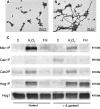
Effects of S. gordonii on C. albicans signaling pathways. (A and B) C. albicans suspensions in YPT-Glc incubated for 4 h at 37°C, showing inhibition of hyphal formation by 30 μM farnesol (A) and suppression of 30 μM farnesol inhibition by S. gordonii DL1 (B). (C) Western immunoblot analysis of effects of 10 mM H2O2 or 0.1 mM farnesol (Fnl) on phosphorylation of C. albicans MAP kinases after 20 min in the absence (left three lanes) or presence (right three lanes) of S. gordonii DL1. MAP kinases were detected with anti-phospho-p44/42 MAP kinase antibody (Mkc1p, Cek1p, Cek2p), anti-phospho-p38 MAP kinase antibody (Hog1p), and an anti-ScHog1 polyclonal antibody (Hog1) control. Experiments were repeated three times, and representative data from one experiment are shown.
FIG. 5.
FIG. 5.
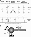
Summary of environmentally responsive signaling pathways and Streptococcus interactive mechanisms in C. albicans. (A) Pathways associated with hyphal development or biofilm formation, based on information from references , , , , and . Various environmental cues (top) interact with cell surface receptors to activate intracellular signaling pathways, leading to transcription factor modulation. cAMP, cyclic AMP; MAPKKK, MAP kinase kinase kinase; MAPKK, MAP kinase kinase; MAPK, MAP kinase; AC, adenylate cyclase; PM, plasma membrane. (B) Proposed interactions of S. gordonii with C. albicans resulting in stimulation of hyphal development and biofilm formation. Streptococcus cells produce diffusible signals (DS) mediated in part through activity of luxS, which might include metabolic by-products, such as H2O2. The effects of these diffusible signals may be intensified by contact signals (CS) generated through attachment of S. gordonii to C. albicans receptors expressed on mother cells or hyphae. Streptococci also appear to suppress the inhibitory effects of farnesol (Fnl) on hyphal formation, which does not involve farnesol inactivation. This may suggest that an alternative signal response generated through recognition of S. gordonii overcomes the repression of morphogenesis as a result of the farnesol-responsive signaling pathway.

References

    1. Alem, M. A., M. D. Oteef, T. H. Flowers, and L. J. Douglas. 2006. Production of tyrosol by Candida albicans biofilms and its role in quorum sensing and biofilm development. Eukaryot. Cell 51770-1779. - PMC - PubMed
    1. Alonso-Monge, R., F. Navarro-Garcia, G. Molero, R. Diez-Orejas, M. Gustin, J. Pla, M. Sanchez, and C. Nombela. 1999. Role of the mitogen-activated protein kinase Hog1p in morphogenesis and virulence of Candida albicans. J. Bacteriol. 1813058-3068. - PMC - PubMed
    1. Bahn, Y. S., M. Molenda, J. F. Staab, C. A. Lyman, L. J. Gordon, and P. Sundstrom. 2007. Genome-wide transcriptional profiling of the cyclic AMP-dependent signaling pathway during morphogenic transitions of Candida albicans. Eukaryot. Cell 62376-2390. - PMC - PubMed
    1. Baillie, G. S., and L. J. Douglas. 1999. Role of dimorphism in the development of Candida albicans biofilms. J. Med. Microbiol. 48671-679. - PubMed
    1. Barnard, J. P., and M. W. Stinson. 1996. The alpha-hemolysin of Streptococcus gordonii is hydrogen peroxide. Infect. Immun. 643853-3857. - PMC - PubMed

Publication types

MeSH terms

Substances

LinkOut - more resources