. 2009 Aug;29(16):4341-51.
doi: 10.1128/MCB.00210-09. Epub 2009 Jun 15.

p38 Mitogen-activated protein kinase- and HuR-dependent stabilization of p21(Cip1) mRNA mediates the G(1)/S checkpoint

Affiliations

p38 Mitogen-activated protein kinase- and HuR-dependent stabilization of p21(Cip1) mRNA mediates the G(1)/S checkpoint

Vanesa Lafarga et al. Mol Cell Biol. 2009 Aug.

Abstract

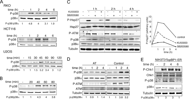
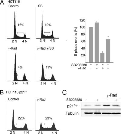
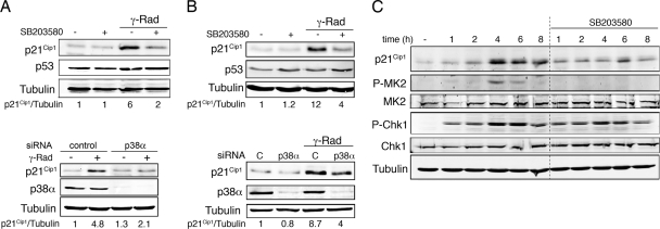
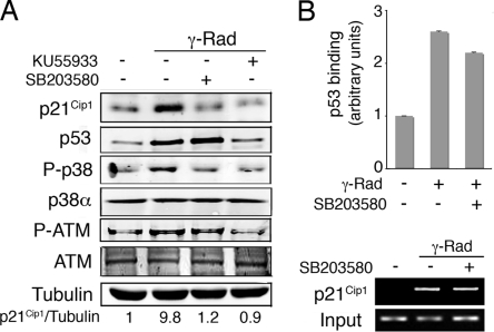
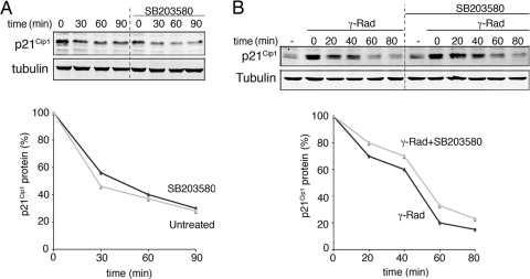
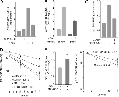
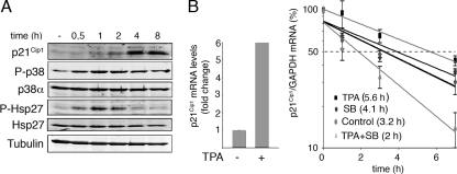
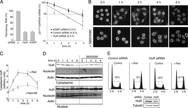
Activation of p38 mitogen-activated protein kinase (MAPK) plays an important role in the G(2)/M cell cycle arrest induced by DNA damage, but little is known about the role of this signaling pathway in the G(1)/S transition. Upregulation of the cyclin-dependent kinase inhibitor p21(Cip1) is thought to make a major contribution to the G(1)/S cell cycle arrest induced by gamma radiation. We show here that inhibition of p38 MAPK impairs p21(Cip1) accumulation and, as a result, the ability of cells to arrest in G(1) in response to gamma radiation. We found that p38 MAPK induces p21(Cip1) mRNA stabilization, without affecting its transcription or the stability of the protein. In particular, p38 MAPK phosphorylates the mRNA binding protein HuR on Thr118, which results in cytoplasmic accumulation of HuR and its enhanced binding to the p21(Cip1) mRNA. Our findings help to understand the emerging role of p38 MAPK in the cellular responses to DNA damage and reveal the existence of p53-independent networks that cooperate in modulating p21(Cip1) levels at the G(1)/S checkpoint.


Figures

FIG. 1.
FIG. 1.
p38α is activated by γ radiation. (A) U2OS and RKO cells were irradiated with 10 Gy and HCT116 cells with 3 Gy, and phospho-p38 levels were analyzed by immunoblotting at the indicated times. (B) U2OS cells were treated with NCS (0.3 μg/ml) and p38 phosphorylation was analyzed by immunoblotting. (C) RKO cells were pretreated for 1 h with KU55933 or 20 min with SB203580 and irradiated with 10 Gy, and the total cell lysates were analyzed by immunoblotting. (D) ATM-deficient (AT) and wt (control) cells were γ irradiated, and p38 phosphorylation was analyzed by immunoblotting at the indicated time points. (E) NIH 3T3 cells expressing TopBP1-ER were incubated with OHT (500 nM) for the indicated times and analyzed by immunoblotting.
FIG. 2.
FIG. 2.
p38α activity is necessary for the establishment of the G1/S checkpoint in response to γ radiation (γ-Rad). HCT116 wt (A) and p21Cip1−/− (B) cells were irradiated with 3 Gy and 16 h later were analyzed by flow cytometry. The percentage of cells in S phase is indicated in the histogram, which shows the average of two independent experiments. (C) Total cell lysates were analyzed by immunoblotting. SB, SB203580.
FIG. 3.
FIG. 3.
γ-radiation-induced p21Cip1 expression depends on p38 MAPK activity. RKO cells (A) and U2OS cells (B) were incubated with SB203580 or were transfected with p38α and control siRNAs before treatment with 10 Gy of γ radiation (γ-Rad). After 16 h, total cell lysates were analyzed by immunoblotting. (C) NIH 3T3 cells expressing TopBP1-ER were treated with OHT alone or together with SB203580 for the indicated times. Total cell lysates were analyzed by immunoblotting.
FIG. 4.
FIG. 4.
p38α regulates p21Cip1 expression levels independently of p53 transcriptional activation. (A) RKO cells were pretreated with KU55933 or SB203580 and then irradiated with 10 Gy. After 6 h, total cell lysates were analyzed by immunoblotting. (B) U2OS cells were incubated in the presence or absence of SB203580 from 30 min before treatment with γ radiation (γ-Rad; 5 Gy). Samples were analyzed by ChIP followed by qRT-PCR (histogram) and semiquantitative RT-PCR (agarose gel).
FIG. 5.
FIG. 5.
p38α does not regulate p21Cip1 protein half-life. (A) RKO cells were pretreated with SB203580 (10 μM) and then incubated with CHX (30 μg/μl) for the indicated times. Total cell lysates were analyzed by immunoblotting. (B) RKO cells were treated with γ radiation (γ-Rad) and 4 h later were processed as in panel A. Quantification of p21Cip1 protein half-life is shown in the graphics below.
FIG. 6.
FIG. 6.
p38α regulates p21Cip1 mRNA half-life. (A and B) RKO cells were treated with SB203580 (SB) or transfected with p38α and control siRNAs and γ irradiated (γ-Rad; 10 Gy), and 4 h later, total RNA was purified. p21Cip1 and GAPDH mRNA levels were analyzed by qRT-PCR. (C) NIH 3T3-TopBP1-ER cells were treated with OHT alone or together with SB203580, and p21Cip1 mRNA levels were measured by qRT-PCR. (D) RKO cells were irradiated (10 Gy) and 3 h later were incubated with actinomycin D in the presence or absence of SB203580 (SB). Total RNA was obtained at the indicated times, and p21Cip1 and GAPDH mRNA levels were analyzed by qRT-PCR. (E) U2OS cells were transfected with vector alone or with both p38α and MKK6DD expression vectors (1 μg each) and 48 h later were treated with actinomycin D for the indicated times. p21Cip1 and GAPDH mRNA levels were measured by qRT-PCR.
FIG. 7.
FIG. 7.
p38 MAPK regulates p21Cip1 mRNA stability independently of p53. (A) HCT116 p53−/− cells were treated with TPA, and total cell lysates were analyzed by immunoblotting at the indicated times. (B) HCT116 p53−/− cells were treated with TPA and 4 h later were incubated with actinomycin D in the presence or absence of SB203580 (SB). Total RNA was obtained at the indicated times, and p21Cip1 and GAPDH mRNA levels were analyzed by qRT-PCR.
FIG. 8.
FIG. 8.
Cytoplasmic localization of HuR after γ radiation (γ-Rad) regulates p21Cip1 mRNA stability. (A) RKO cells were transfected with HuR, KSRP, or control (C) siRNAs and 48 h later were γ irradiated with 10 Gy for 4 h and then treated with actinomycin D for the indicated times. Total RNA was obtained at the indicated times, and mRNA levels were analyzed by qRT-PCR. (B) RKO cells were treated with γ radiation (10 Gy) in the presence or absence of SB203580, and HuR localization was visualized by confocal microscopy at the indicated times after irradiation. Bar, 5 μm. (C) Quantification of fluorescence intensities for the cytosolic HuR of the samples in panel B obtained using a confocal laser microscope and the Analysis of Image system. (D) Immunoblotting analysis of HuR levels in cytoplasmic and nuclear fractions of RKO cells at the indicated times after γ radiation (10 Gy). Antibodies against actin and nucleolin were used to confirm equal loading of the cytoplasmic and nuclear samples, respectively. (E) U2OS cells were transfected with HuR and control siRNAs and 48 h later were treated with 3 Gy of γ radiation, incubated for 16 h, and analyzed by flow cytometry. The immunoblot shows HuR protein expression.
FIG. 9.
FIG. 9.
p38α phosphorylates HuR on Thr118. (A) Kinase assays were performed in the presence of [γ-32P]ATP using MKK6DD-activated GST-p38α (200 ng) and GST, GST-HuR wt or GST-HuR-T118A as substrates (1 μg). The gels were stained with Coomassie blue and then exposed to autoradiography. (B) GST-HuR wt and GST-HuR-T118A proteins (500 ng) were incubated with MKK6DD-activated GST-p38α and then analyzed by immunoblotting with a phospho-Thr antibody. (C) RKO cells were transfected with HA-tagged HuR wt or mutant T118A and MKK6DD plus p38α, as indicated. After 48 h, HuR phosphorylation was analyzed by HA IP followed by immunoblotting with a phospho-Thr antibody. Total cell lysates were also analyzed by immunoblotting. (D) U2OS cells were transfected with HA-HuR wt or T118A and 24 h later were treated with γ radiation. HuR localization was visualized by confocal microscopy at the indicated times after γ radiation using an HA antibody to avoid the endogenous HuR protein. Bar, 5 μm. The percentage of cells (out of 300 counted) with a cytoplasmic HuR signal is shown in the right panel. (E) U2OS cells were transfected with p38α or HuR siRNAs and 48 h later were transfected again with HA-tagged HuR wt or the T118A mutant. Total cell lysates were analyzed by immunoblotting.
FIG. 10.
FIG. 10.
γ radiation induces the binding of HuR to p21Cip1 mRNA. (A) RKO cells were γ irradiated (γ-Rad; 10 Gy) and treated with SB203580, as indicated, and total cell lysates were then immunoprecipitated with either HuR antibodies or control IgGs. RNA was purified from the immunoprecipitates and used for qRT-PCRs. The graph shows fold differences in transcript abundance in HuR immunoprecipitates compared with the IgG immunoprecipitates. The image shows a representative PCR visualized by Syber-safe staining of the agarose gel. (B) RKO cells were transfected with constructs encoding HA-tagged HuR wt and T118A mutant proteins, and total cell lysates were then immunoprecipitated with HA antibody. RNA was purified from the immunoprecipitates, and the amount of p21Cip1 mRNA was determined by qRT-PCR. The expression levels of the different HA-HuR proteins were analyzed by immunoblotting. (C) A model for the p38 MAPK-dependent upregulation of p21Cip1 in response to γ radiation.

Similar articles

Cited by

References

    1. Abdelmohsen, K., R. Pullmann, Jr., A. Lal, H. H. Kim, S. Galban, X. Yang, J. D. Blethrow, M. Walker, J. Shubert, D. A. Gillespie, H. Furneaux, and M. Gorospe. 2007. Phosphorylation of HuR by Chk2 regulates SIRT1 expression. Mol. Cell 25543-557. - PMC - PubMed
    1. Abdelmohsen, K., S. Srikantan, X. Yang, A. Lal, H. H. Kim, Y. Kuwano, S. Galban, K. G. Becker, D. Kamara, R. de Cabo, and M. Gorospe. 2009. Ubiquitin-mediated proteolysis of HuR by heat shock. EMBO J. 281271-1282. - PMC - PubMed
    1. Alonso, G., C. Ambrosino, M. Jones, and A. R. Nebreda. 2000. Differential activation of p38 mitogen-activated protein kinase isoforms depending on signal strength. J. Biol. Chem. 27540641-40648. - PubMed
    1. Ambrosino, C., G. Mace, S. Galban, C. Fritsch, K. Vintersten, E. Black, M. Gorospe, and A. R. Nebreda. 2003. Negative feedback regulation of MKK6 mRNA stability by p38α mitogen-activated protein kinase. Mol. Cell. Biol. 23370-381. - PMC - PubMed
    1. Bakkenist, C. J., and M. B. Kastan. 2003. DNA damage activates ATM through intermolecular autophosphorylation and dimer dissociation. Nature 421499-506. - PubMed

Publication types

MeSH terms