

Pregnancy and estrogen receptor beta expression in a large congenital nevus
- PMID: 19528425
- PMCID: PMC3860585
- DOI: 10.1001/archdermatol.2009.72
Pregnancy and estrogen receptor beta expression in a large congenital nevus
Abstract
Background: Large congenital nevi carry a slightly increased risk of melanoma. Pregnancy poses an additional challenge in the monitoring of these patients because little is known regarding the effects of increased estrogen levels on congenital nevi.
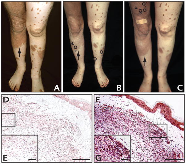
Observations: A young woman was observed to have clinical lightening of her garment nevus and satellite nevi during 2 sequential pregnancies. Postpartum, the patient experienced darkening and repigmentation in her large garment nevus, with continued lightening of nearby satellite lesions. In addition to photographic documentation of these changes, biopsy samples taken during pregnant and nonpregnant periods underwent immunohistochemical evaluation for estrogen receptor beta (ERbeta), the predominant estrogen receptor in nevi and melanomas. Biopsy samples collected during pregnancy showed a decrease in nuclear staining for ERbeta compared with samples collected after pregnancy. These changes in ERbeta expression were not associated with histologic atypia during pregnancy or after delivery.
Conclusions: Congenital nevi may be unique in their response to altered estrogen levels. Given the slightly increased risk of melanoma in giant congenital nevi and the dearth of information available regarding the effects of pregnancy on congenital nevi, this case illustrates the need for further study of these pigmented lesions.
Figures
References
-
- Bett BJ. Large or multiple congenital melanocytic nevi: occurrence of cutaneous melanoma in 1008 persons. J Am Acad Dermatol. 2005;52:793–797. - PubMed
-
- Ellis DL, Wheeland RG, Solomon H. Estrogen and progesterone receptors in congenital melanocytic nevi. J Am Acad Dermatol. 1985;12:235–244. - PubMed
-
- Hu W, Nelson JE, Mohney CA, Willen MD. Malignant melanoma arising in a pregnant African American woman with a congenital blue nevus. Dermatol Surg. 2004;30:1530–1532. - PubMed
-
- Streams BN, Lio PA, Mihm MC, Sober AJ. A nonepidermal, primary malignant melanoma arising in a giant congenital melanocytic nevus 40 years after partial surgical removal. J Am Acad Dermatol. 2004;50:789–792. - PubMed
-
- Swerdlow AJ, English JS, Qiao Z. The risk of melanoma in patients with congenital nevi: a cohort study. J Am Acad Dermatol. 1995;32:595–599. - PubMed
Publication types
MeSH terms
Substances
Grants and funding
LinkOut - more resources
Full Text Sources
Medical
