Review
. 2009 Nov;19(11):1155-62.
doi: 10.1093/glycob/cwp076. Epub 2009 Jun 15.

Structural insights into what glycan arrays tell us about how glycan-binding proteins interact with their ligands

Affiliations
Review

Structural insights into what glycan arrays tell us about how glycan-binding proteins interact with their ligands

Maureen E Taylor et al. Glycobiology. 2009 Nov.

Abstract

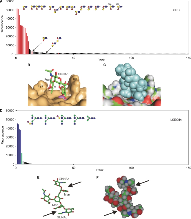
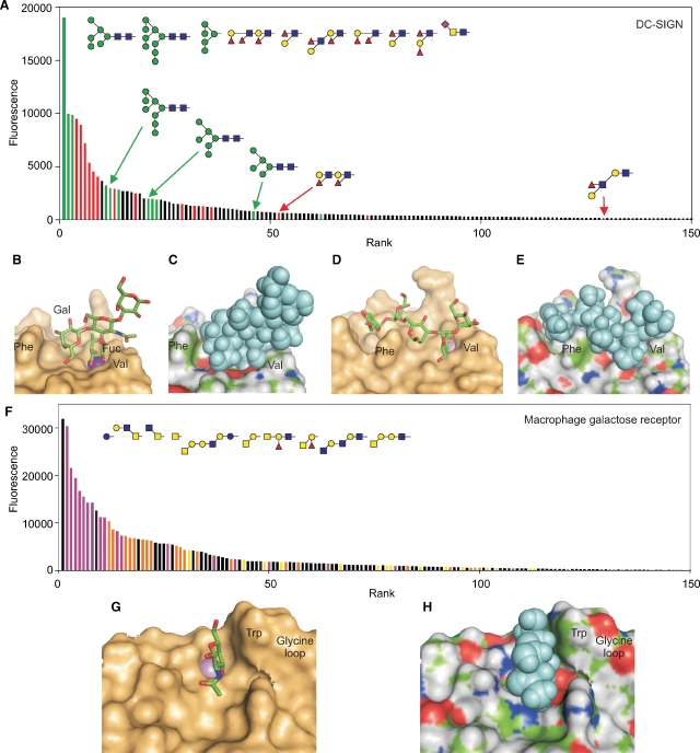
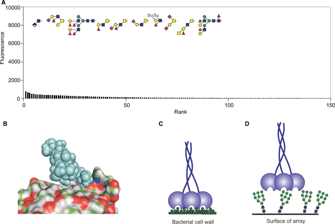
Screening of glycan arrays represents a powerful, high-throughput approach to defining oligosaccharide ligands for glycan-binding receptors, commonly referred to as lectins. Correlating results from such arrays with structural analysis of receptor-ligand complexes provide one way to validate the arrays. Using examples drawn from the family of proteins that contain C-type carbohydrate-recognition domains, this review illustrates how information from the arrays reflects the way that selectivity and affinity for glycan ligands is achieved. A range of binding profiles is observed, from very restricted binding to a small set of structurally similar ligands to binding of broad classes of ligands with related terminal sugars and even to failure to bind any of the glycans on an array. These outcomes provide insights into the importance of multiple factors in defining the selectivity of these receptors, including the presence of conformationally defined units in some oligosaccharide ligands, local and extended interactions between glycans and the surfaces of receptors, and steric factors that exclude binding of some ligands.


Figures

Fig. 1
Fig. 1
Ligand binding by the scavenger receptor C-type lectin (SRCL) and LSECtin. (A) Glycan array data for the mouse receptor (200 μg/mL) plotted in rank order of ligand binding. The 10 oligosaccharides giving the highest signals are highlighted in red, along with other structurally similar glycans that are ranked lower. (B) Structure of Lewisx trisaccharide bound to SRCL. The surface of the protein is shown, with Ca2+ highlighted in violet. The A face of fucose and the A and B faces of galactose are labeled. These designations are based on the order of the ring carbons, which is clockwise when the sugar is viewed from the A face. (C) Structure of the complex in (B) with surface colored by underlying atom type and ligand presented as space-filling spheres. (D) Results for glycan arrays probed with labeled human LSECtin CRD-streptavidin tetramers (4.5 μg/mL). The structures of the five oligosaccharides giving the highest signals are shown. Bars are coded based on terminal sugars in the glycans: green for mannose and blue for GlcNAc. (E and F) Structure of a portion of a bi-antennary glycan terminating in GlcNAc-β1-2Man disaccharides showing packing of the N-acetyl group of GlcNAc on top of mannose. The figure was created from CFG primscreen_GLYCAN_v3_72_02172005 and CFG primscreen_1043 and structures 2OX9 and 1SLA in the PDB.
Fig. 2
Fig. 2
Binding of multiple classes of ligands to DC-SIGN and the macrophage galactose receptor. (A) Data for screening of a recent version of the Consortium for Functional Glycomics glycan array with human DC-SIGN (200 μg/mL). Results are presented in rank order of binding, with bars for mannose-terminated glycans colored green and bars for glycans containing the Lewisa or Lewisx epitopes highlighted in red. The 10 oligosaccharides giving the strongest signals are shown at the top and additional related glycans are indicated below. (B and C) Structure of the CRD from DC-SIGN in complex with a tetrasaccharide containing the Lewisx epitope. (D and E) Structure of the CRD with Man5 modeled into the binding site based on the crystal structure with a GlcNAc2Man3 oligosaccharide. (F) Binding data for the human macrophage galactose receptor (4.5 μg/mL) on the CFG printed glycan array. The results are similar to previous studies with the human and rat receptors on earlier streptavidin-based arrays (Coombs et al. ; van Vliet et al. 2005). The data are color-coded to indicate glycans bearing GalNAc with free 3 and 4 hydroxyl groups in purple and galactose with free 3 and 4 hydroxyl groups in orange, except galactose residues in α linkage or adjacent to fucose residues, which are in yellow. In spite of the preference for exposed GalNAc and galactose residues, binding of other glycans indicated in black also occurs, including the simple sugar glucose which gives the highest signal. (G and H) Model of the binding site in the macrophage galactose receptor with a bound GalNAc residue, based on the structure of the galactose-binding mutant of mannose-binding protein that was created by insertion of key binding site residues from the galactose-binding receptor. These residues include the marked tryptophan residue and the glycine-rich loop positioned just to the right of this residue, which serves to hold it in the position for packing against galactose or GalNAc residues. For the model, additional surface residues were substituted into the structure using Insight software. Data are from CFG primscreen_2010, and the model structures were created starting from entries 1sl5, 1k9i, and 1afb in the PDB.
Fig. 3
Fig. 3
Mechanisms of mannose-binding protein interaction with ligands. (A) Screening of the glycan array with the trimeric terminal fragment of rat serum mannose-binding protein at a 250 μg/mL concentration. The oligosaccharides giving the 10 highest signals are illustrated at the top. Scale of the y-axis is expanded compared to other figures. (B) Structure of a Man6 ligand complexed with the CRD of mannose-binding protein. (C and D) Diagrams illustrating the potential effect of glycan spacing on the avidity with which mannose-binding protein trimers bind to dense clusters of sugars on bacterial or fungal surfaces compared to their interaction with glycans on the array. The figure is based on data from CFG primscreen_1618 and structure 1kx1 in the PDB.

References

    1. Blixt O, Head S, Mondala T, Scanlan C, Huflejt ME, Alvarez R, Bryan MC, Fazio F, Calarese D, Stevens J, et al. Printed covalent glycan array for ligand profiling of diverse glycan binding proteins. Proc Natl Acad Sci USA. 2004;101:17033–17038. - PMC - PubMed
    1. Bochner BS, Alvarez RA, Mehta P, Bovin NV, Blixt O, White JR, Schnaar RL. Glycan array screening reveals a candidate ligand for siglec-8. J Biol Chem. 2005;280:4307–4312. - PubMed
    1. Coombs PJ, Graham SA, Drickamer K, Taylor ME. Selective binding of the scavenger receptor C-type lectin to Lewisx trisaccharide and related glycan ligands. J Biol Chem. 2005;280:22993–22999. - PubMed
    1. Engering A, Geijtenbeek TBH, van Vliet SJ, Wijers M, van Liempt E, Demaurex N, Lanzacecchia A, Fransen J, Figdor CG, Piguet V, et al. The dendritic cell-specific adhesion receptor DC-SIGN internalizes antigen for presentation to T cells. J Immunol. 2002;168:2118–2126. - PubMed
    1. Feinberg H, Castelli R, Drickamer K, Seeberger PH, Weis WI. Multiple modes of binding enhance the affinity of DC-SIGN for high-mannose N-linked glycans found on viral glycoproteins. J Biol Chem. 2007;282:4202–4209. - PMC - PubMed

Publication types