. 2009;4(5):767-82.
doi: 10.1038/nprot.2009.40.

Screening individual hybridomas by microengraving to discover monoclonal antibodies

Affiliations

Screening individual hybridomas by microengraving to discover monoclonal antibodies

Adebola O Ogunniyi et al. Nat Protoc. 2009.

Abstract

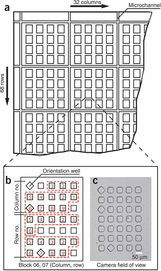
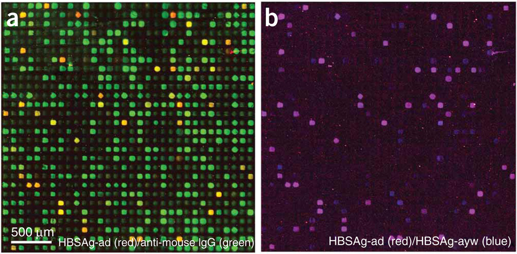
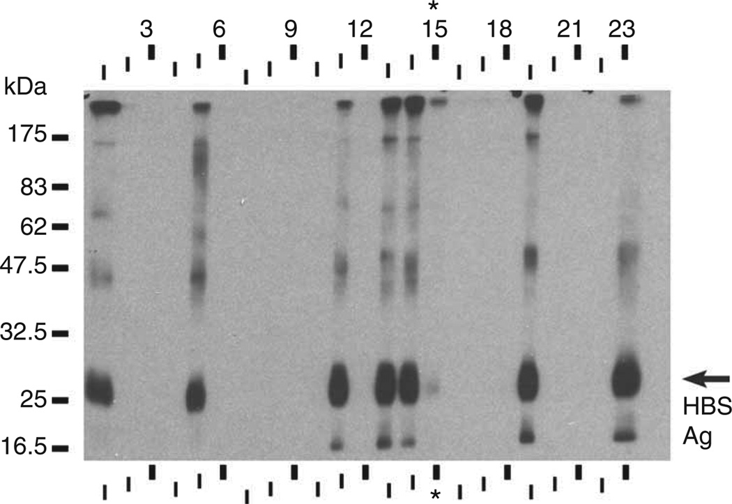
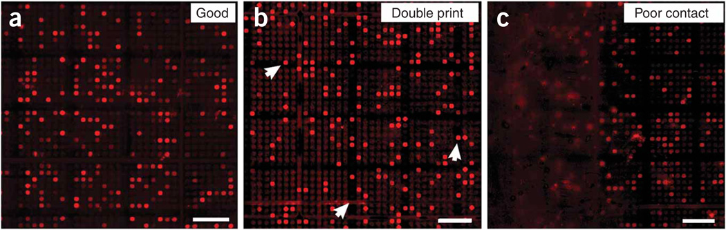
The demand for monoclonal antibodies (mAbs) in biomedical research is significant, but the current methodologies used to discover them are both lengthy and costly. Consequently, the diversity of antibodies available for any particular antigen remains limited. Microengraving is a soft lithographic technique that provides a rapid and efficient alternative for discovering new mAbs. This protocol describes how to use microengraving to screen mouse hybridomas to establish new cell lines producing unique mAbs. Single cells from a polyclonal population are isolated into an array of microscale wells (approximately 10(5) cells per screen). The array is then used to print a protein microarray, where each element contains the antibodies captured from individual wells. The antibodies on the microarray are screened with antigens of interest, and mapped to the corresponding cells, which are then recovered from their microwells by micromanipulation. Screening and retrieval require approximately 1-3 d (9-12 d including the steps for preparing arrays of microwells).


Figures

Figure 1
Figure 1
Schematic diagram of the processes described in this protocol. Steps shown parallel to one another can be carried out concurrently.
Figure 2
Figure 2
Design of an array of microwells for microengraving. (a) Schematic representation of the top-left corner of the array showing a network of microchannels and 4 × 4 groups of blocks containing microwells. (b) Annotated illustration of one block comprising a 5 × 7 arrangement of microwells. Each well projects a square cross-section and has a cuboid geometry in the molded array. Certain wells are rotated 45° within a given block to encode the precise position of that block within the array of blocks (e.g., 32 × 68). The rotated well in the top-left corner of a block establishes the orientation of the array and is present in the same position in every block. The presence or absence of rotated wells in other areas of the block encodes the location (row and column) of the block within the entire array. Here, the areas outlined with red, dashed lines indicate the block column and row numbers as two-digit integers (columns 01–32, rows 01–68). In the area labeled on the scheme for encoding the column, the topmost region (bound by red, dashed lines) is used to indicate the first digit of the column number, whereas the region immediately below indicates the second digit of column number. The same scheme is applied in the lower region of the block to encode the row. The position of a rotated well within these designated areas specifies the first or second digit of the column or row number; zero is indicated by the absence of a rotated well. In the example shown here, the positions of the rotated wells, read from top to bottom, indicate Block 06, 07 (column, row). (c) Transmitted-light micrograph of Block 06, 07 loaded with hybridoma cells (HYB9901) acquired at a magnification of ×10 with a Hamamatsu ORCA-AG digital camera. The entire field of view of the camera is shown. Scale bar, 50 µm.
Figure 3
Figure 3
Retrieval of cells from microwells with a micromanipulator. (a) Phase-contrast image of a section of a microwell array with a 20-µm-diameter GC-1 capillary tip (Narishige), attached to an IM-9A micromanipulator (Narishige), inserted into a well loaded with cells. (b) Phase-contrast image of the microwell array section in a, with the cells selectively removed from the central microwell. (c) The cells recovered from the central microwell in a were transferred into a well of a 96-well plate containing cloning media, and cultured until confluent. Note that the design of the array of microwells used here differs from the example shown in Figure 2. Scale bar, 100 µm.
Figure 4
Figure 4
Microarrays of antibodies produced by microengraving. The source of cells was a polyclonal population of murine hybridomas generated from a mouse immunized with hepatitis B antigens, HBSAg-ayw and HBSAg-ad. (a) Composite fluorescence micrograph showing signals from both HBSAg-ad (red) and IgG (green). HBSAg-ad-specific antibodies appear yellow in the micrograph. (b) Composite fluorescence micrograph showing subset of antibodies in a, with specificity to HBSAg-ad (red) and HBSAg-ayw (blue). Antibodies that bind epitopes on both HBSAg-ad and HBSAg-ayw appear purple in the micrograph.
Figure 5
Figure 5
‘Curtain’ western blot of 12.5% polyacrylamide gel loaded with 200 µg of complete COS-1 cell lysate with 10 µg HBSAg (ViroStat). Using a Surfblot device (Idea Scientific), a different culture supernatant was applied to each strip, numbered 1–23, except lane 15 to which sera from the immunized mouse (1:200 dilution) was applied (asterisk). The film for the blot shown was exposed for 60 s. A duplicate blot without added HBSAg was blank (data not shown).
Figure 6
Figure 6
Examples of potential variations in the quality of the microarrays produced by microengraving. (a) Micrograph of a typical, high-quality microarray resulting from successful application of the technique. The position of each positive spot is unique, and each spot has a well-defined shape. (b) Micrograph from a microarray obtained from a print in which the array of microwells moved while sealing to or separating from the glass slide. Double printing will occur if the array is lifted and repositioned, dragged over the surface of the slide, or if uneven pressure is applied to PDMS; the latter two may also result in smeared features. Arrowheads highlight some features that are out of register. (c) Micrograph of a microarray resulting from poor contact between the glass slide and the array of microwells. Secreted antibodies diffused over the surface of the glass, and individual, localized features are not evident. The contrast and brightness of each image was optimized automatically by GenePix Pro. Note that the format of the arrays in these images differs slightly from the scheme shown in Figure 2; the blocks in these arrays comprised 7 × 7 microwells. Scale bars, 500 µm.

References

    1. Barrett W, Newsome MSE. The clinical pharmacology of therapeutic monoclonal antibodies in the treatment of malignancy; have the magic bullets arrived? Br. J. Clin. Pharmacol. 2008;66:6–19. - PMC - PubMed
    1. Finn OJ. Molecular origins of cancer—cancer immunology. N. Engl. J. Med. 2008;358:2704–2715. - PubMed
    1. Buttmann M, Rieckmann P. Treating multiple sclerosis with monoclonal antibodies. Exp. Rev. Neurotherapeutics. 2008;8:433–455. - PubMed
    1. Jennifer MD, Stuart ML. Prospects for development of vaccines against fungal diseases. Drug Resist. Updat. 2006;9:105–110. - PubMed
    1. Keller MA, Stiehm ER. Passive immunity in prevention and treatment of infectious diseases. Clin. Microbiol. Rev. 2000;13:602–614. - PMC - PubMed

Publication types

Substances