. 2009 Jun 16;4(6):e5930.
doi: 10.1371/journal.pone.0005930.

Protection and polyfunctional T cells induced by Ag85B-TB10.4/IC31 against Mycobacterium tuberculosis is highly dependent on the antigen dose

Affiliations

Protection and polyfunctional T cells induced by Ag85B-TB10.4/IC31 against Mycobacterium tuberculosis is highly dependent on the antigen dose

Claus Aagaard et al. PLoS One. .

Abstract

Background: Previously we have shown that Ag85B-TB10.4 is a highly efficient vaccine against tuberculosis when delivered in a Th1 inducing adjuvant based on cationic liposomes. Another Th1 inducing adjuvant, which has shown a very promising profile in both preclinical and clinical trials, is IC31. In this study, we examined the potential of Ag85B-TB10.4 delivered in the adjuvant IC31 for the ability to induce protection against infection with Mycobacterium tuberculosis. In addition, we examined if the antigen dose could influence the phenotype of the induced T cells.

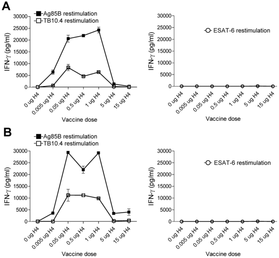
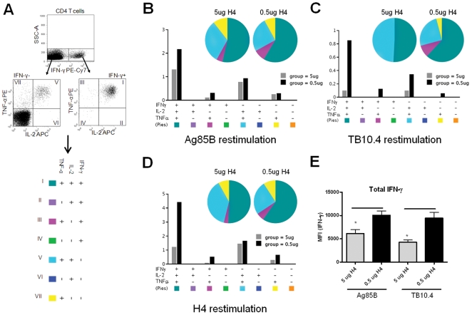
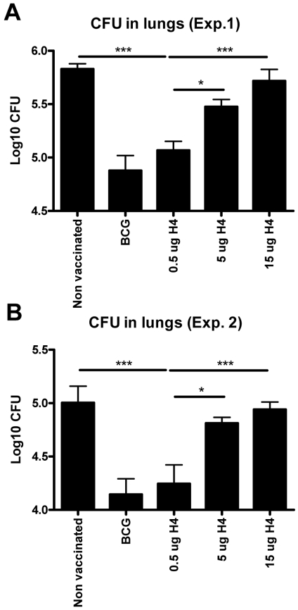
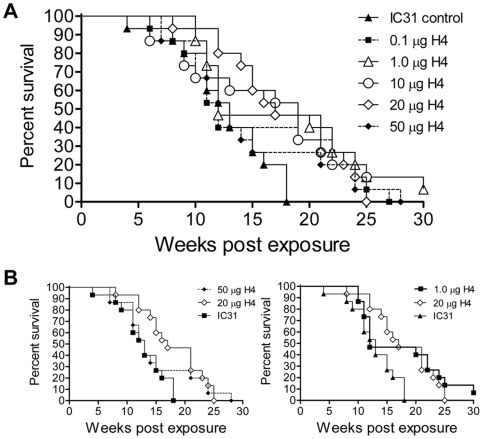
Methods and findings: We found that vaccination with the combination of Ag85B-TB10.4 and IC31 resulted in high numbers of polyfunctional CD4 T cells co-expressing IL-2, IFN-gamma and TNF-alpha. This correlated with protection against subsequent challenge with M.tb in the mouse TB model. Importantly, our results also showed that both the vaccine induced T cell response, and the protective efficacy, was highly dependent on the antigen dose. Thus, whereas antigen doses of 5 and 15 microg did not induce significant protection against M.tb, reducing the dose to 0.5 microg selectively increased the number of polyfunctional T cells and induced a strong protection against infection with M.tb. The influence of antigen dose was also observed in the guinea pig model of aerosol infection with M.tb. In this model a 2.5 fold increase in the antigen dose reduced the protection against infection with M.tb to the level observed in non-vaccinated animals.

Conclusions/significance: Small changes in the antigen dose can greatly influence the induction of specific T cell subpopulations and the dose is therefore a crucial factor when testing new vaccines. However, the adjuvant IC31 can, with the optimal dose of Ag85B-TB10.4, induce strong protection against Mycobacterium tuberculosis. This vaccine has now entered clinical trials.


Conflict of interest statement

Competing Interests: The authors have declared that no competing interests exist.

Figures

Figure 1
Figure 1. Immune recognition of vaccination antigens.
PBMC's (A) or splenocytes (B) isolated from groups of mice vaccinated with different doses of Ag85B-TB10.4 in IC31® (and a saline control group) were stimulated with either Ag85B or TB10.4 (or ESAT-6 as negative control). After 72 hours the concentration of cell released IFN-γ was determined by ELISA. Five mice per group were pooled. Values represent the means of triplicate and SEM's are indicated.
Figure 2
Figure 2. ELISPOT analysis of splenocytes from vaccinated animals.
(A and B) Splenocytes isolated from groups of mice vaccinated with 3 different doses of Ag85B-TB10.4 in IC31® or a saline control group (Non Vacc.) were stimulated in vitro with Ag85B for 48 hours and subjected to ELISPOT analysis (A) or stimulated for 72 hours to measure IFN-γ cytokine secretion by ELISA (B). The bars represent means of 3 individual mice. SEMs are indicated. In both (A) and (B) a vaccination dose of 0.5 µg Ag85B-TB10.4 gave significantly (*p<0.05, one-way ANOVA and Tukey's post test) higher antigen responses, compared to vaccination doses of 5 and 15 µg.
Figure 3
Figure 3. Ag85B and TB10.4 specific T cells are polyfunctional.
(A). Cytokine profiles of H4 specific CD4 T cells were determined by first dividing the CD4 T cells into IFN-γ positive (+) or IFN-γ negative (-) cells. Both the IFN-γ+ and IFN-γ cells were analyzed with respect to the production of TNF-α and IL-2. The numbers in the quadrant gates of the plots denominates each distinct population based on their cytokine production and is color coded as shown. (B–D) The pie charts are grouped after vaccination dose and colour coded according to the cytokine production profile and summarizes the fractions of the CD4+ T cell response (out of the antigen specific CD4 T cells) that are positive for a given cytokine production profile. Every possible combination of cytokines is shown on the x-axis of the bar chart and the percentage of Ag85B, TB10.4 or Ag85B-TB10.4 (H4) specific CD4+ T cells expressing any combination of cytokines is given for each immunization group. The antigen used for in vitro stimulation of the PBMC's is indicated. No responses were seen in the CD8+ T cell subset. (E) The mean fluorescence intensity (MFI) of IFN-γ in the subpopulations expressing this cytokine from animals vaccinated with either 0.5 µg H4 or 5 µg H4. Results are representative of two independent experiments.
Figure 4
Figure 4. Protective efficacy of different doses of Ag85B-TB10.4 in IC31®.
In two independent experiments (A and B) groups of mice were vaccinated with three different doses of H4 formulated in IC31® and compared to saline and BCG vaccinated controls. All groups were challenged by the aerosol route with virulent M.tb ten weeks after the first vaccination. Six weeks post-challenge, all mice were killed and the bacterial burden (CFU) was measured in the lung. In both experiments data are presented as mean values from six animals per group and standard errors of the means are indicated by bars. Statistical comparison among the vaccination groups were done by one-way ANOVA and Tukey's post test. Significant differences are shown. ***: p<0.001, *: p<0.05.
Figure 5
Figure 5. Kaplan-Meier plot of different doses of Ag85B-TB10.4 in the guinea pig TB model.
(A and B). Groups of 15 animals per group were vaccinated three times with Ag85B-TB10.4 in IC31® or with IC31® alone (as a negative control). For antigen dose we used from 0.1 µg to 50 µg. 10 weeks after the last vaccination the animals were infected via the aerosol route with virulent M. tuberculosis, and monitored for weight loss post infection. B. Selected groups from the same experiment.

References

    1. Horwitz MA, Harth G, Dillon BJ, Maslesa-Galic S. Recombinant bacillus calmette-guerin (BCG) vaccines expressing the Mycobacterium tuberculosis 30-kDa major secretory protein induce greater protective immunity against tuberculosis than conventional BCG vaccines in a highly susceptible animal model. Proc Natl Acad Sci U S A. 2000;97:13853–13858. - PMC - PubMed
    1. McShane H, Pathan AA, Sander CR, Keating SM, Gilbert SC, et al. Recombinant modified vaccinia virus Ankara expressing antigen 85A boosts BCG-primed and naturally acquired antimycobacterial immunity in humans. Nat Med. 2004;10:1240–1244. - PubMed
    1. Skeiky YA, Alderson MR, Ovendale PJ, Guderian JA, Brandt L, et al. Differential immune responses and protective efficacy induced by components of a tuberculosis polyprotein vaccine, Mtb72F, delivered as naked DNA or recombinant protein. J Immunol. 2004;172:7618–7628. - PubMed
    1. Olsen AW, van Pinxteren LA, Okkels LM, Rasmussen PB, Andersen P. Protection of mice with a tuberculosis subunit vaccine based on a fusion protein of antigen 85b and esat-6. Infection and Immunity. 2001;69:2773–2778. - PMC - PubMed
    1. Dietrich J, Andersen C, Rappuoli R, Doherty TM, Jensen CG, et al. Mucosal administration of Ag85B-ESAT-6 protects against infection with Mycobacterium tuberculosis and boosts prior bacillus Calmette-Guerin immunity. J Immunol. 2006;177:6353–6360. - PubMed

Publication types

MeSH terms