Case Reports
. 2009 Jul;149A(7):1382-91.
doi: 10.1002/ajmg.a.32846.

Review of disrupted sleep patterns in Smith-Magenis syndrome and normal melatonin secretion in a patient with an atypical interstitial 17p11.2 deletion

Affiliations
Case Reports

Review of disrupted sleep patterns in Smith-Magenis syndrome and normal melatonin secretion in a patient with an atypical interstitial 17p11.2 deletion

Eilis A Boudreau et al. Am J Med Genet A. 2009 Jul.

Abstract

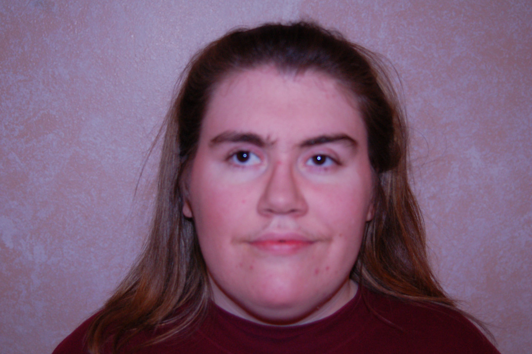
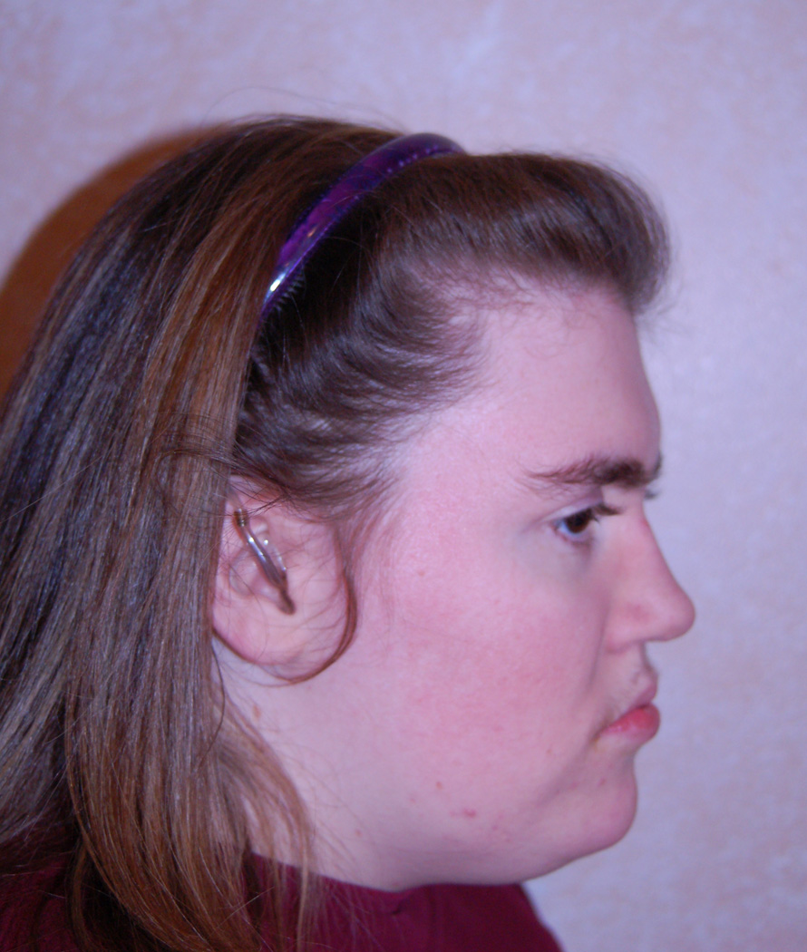
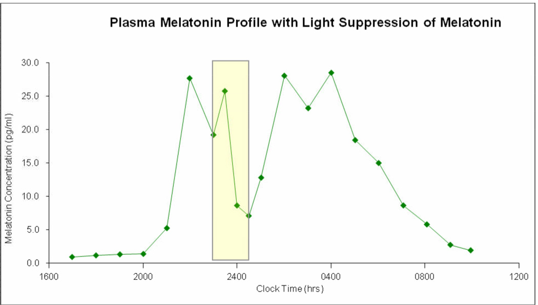
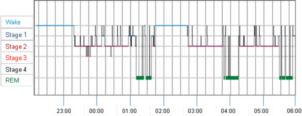
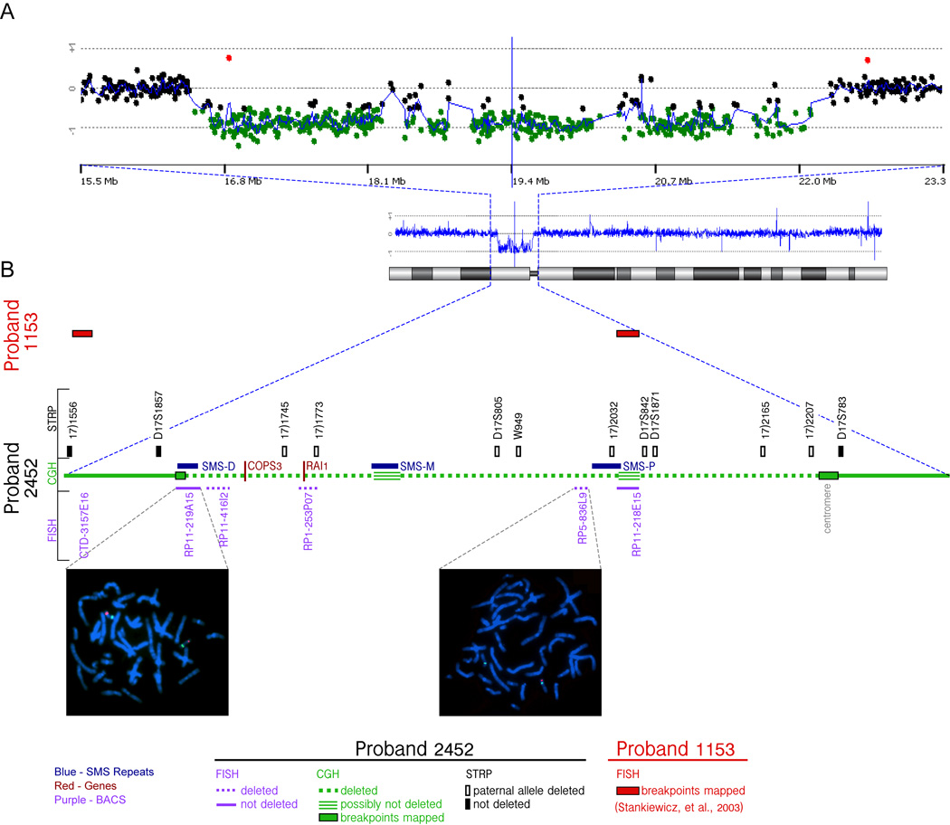
Smith-Magenis syndrome (SMS) is a disorder characterized by multiple congenital anomalies and behavior problems, including abnormal sleep patterns. It is most commonly due to a 3.5 Mb interstitial deletion of chromosome 17 band p11.2. Secretion of melatonin, a hormone produced by the pineal gland, is the body's signal for nighttime darkness. Published reports of 24-hr melatonin secretion patterns in two independent SMS cohorts (US and France) document an inverted endogenous melatonin pattern in virtually all cases (96%), suggesting that this finding is pathognomic for the syndrome. We report on a woman with SMS due to an atypical large proximal deletion ( approximately 6Mb; cen<->TNFRSFproteinB) of chromosome band (17)(p11.2p11.2) who presents with typical sleep disturbances but a normal pattern of melatonin secretion. We further describe a melatonin light suppression test in this patient. This is the second reported patient with a normal endogenous melatonin rhythm in SMS associated with an atypical large deletion. These two patients are significant because they suggest that the sleep disturbances in SMS cannot be solely attributed to the abnormal diurnal melatonin secretion versus the normal nocturnal pattern.


Figures

Figure 1
Figure 1
Photograph of patient showing the typical facial appearance (a. full face and b. profile) seen in SMS including broad forehead, low frontal hairline, square-shaped face, deep-set eyes with upslanting palpebral fissures, short upturned nose, short philtrum, open mouth posture, and maxillary hypoplasia with relative prognathism.
Figure 1
Figure 1
Photograph of patient showing the typical facial appearance (a. full face and b. profile) seen in SMS including broad forehead, low frontal hairline, square-shaped face, deep-set eyes with upslanting palpebral fissures, short upturned nose, short philtrum, open mouth posture, and maxillary hypoplasia with relative prognathism.
Figure 2
Figure 2
Plot of clock time (x-axis) versus plasma melatonin concentration (y-axis) showing the normal pattern of melatonin rise in the evening and nadir in the middle of the day for our patient at age 18 years old. This profile was obtained on the third admission and is nearly identical to those obtained on the first two admissions. It illustrates the peak onset of melatonin occurs at about 2200 hours. It also shows that giving a pulse of bright light (indicated by the rectangles) temporarily inhibits melatonin secretion as shown by the drop in melatonin concentration, which rebounds after the light is stopped. This pattern of inhibition of melatonin secretion by light is typical and further illustrates that melatonin secretion and response to light is not abnormal in our patient.
Figure 3
Figure 3
Hypnogram showing clock time plotted on the x-axis versus sleep stage on the y-axis taken from standard overnight polysomnography on our patient. This figure demonstrates that compared with normative data [Lavie, 2001; Wilson and Nutt, 2008] the patient had increased sleep fragmentation and an extended period of wakefulness between 01:45 and 02:45 AM.
Figure 4
Figure 4
Molecular cytogenetic characterization of the deletion in the patient (2452). A) CGH analysis of chromosome 17 for the proband 2452. Ideogram of the chromosome (below), and the CGH profile scaled to the whole chromosome(middle) is shown along with expansion (top) of the deleted area. The zoomed area (15.5 Mb to 23.3 Mb) includes black spots corresponding to the array CGH probes with “balanced” ratio between control and patient DNA, and green spots showing lower ratio corresponding to the “deleted” region. A CGH profile of the whole chromosome (blue line) gives the mean value of the CGH ratios for 5 consecutive spots. B) The STRP analysis and FISH mapping of the region for the patient 2452 are shown adjacent to the green line representing CGH data. The loss of the paternal alleles (open rectangles) or the presence of both alleles (filled rectangles) for the informative STRP markers are shown. The FISH signals (red) for the BAC RP11-219A15 shows the retention of two copies whereas that for the BAC RP5-836L9 shows deletion of a copy: Green signals indicate a reference probe on 17q or at centromere, respectively. The two BAC clones, CTD-3157E16 and RP11-218E15, in which the distal and proximal breakpoints for the patient 1153 are reported to reside, are indicated (red rectangles) [Stankiewicz et al., 2003]. The sequence coordinates of all the elements shown in this figure, along with the sequence and allele sizes of the STRP markers in the proband 2452 is shown in Supplementary Table I.

References

    1. Benson G. Tandem repeats finder: a program to analyze DNA sequences. Nucleic Acids Res. 1999;27(2):573–580. - PMC - PubMed
    1. Bi W, Saifi GM, Girirajan S, Shi X, Szomju B, Firth H, Magenis RE, Potocki L, Elsea SH, Lupski JR. RAI1 point mutations, CAG repeat variation, and SNP analysis in non-deletion Smith-Magenis syndrome. Am J Med Genet A. 2006;140(22):2454–2463. - PubMed
    1. Cornelissen G, Halberg F, Tarquini R, Perfetto F, Salti R, Laffi G, Otsuka K. Point and interval estimations of circadian melatonin ecphasia in Smith-Magenis syndrome. Biomed Pharmacother. 2003;57 Suppl 1:31s–34s. - PubMed
    1. Czeisler CA, Shanahan TL, Klerman EB, Martens H, Brotman DJ, Emens JS, Klein T, Rizzo JF. Suppression of Melatonin Secretion in Some Blind Patients by Exposure to Bright Light. N Engl J Med. 1995;332(1):6–11. - PubMed
    1. Danel T, Touitou Y. Alcohol Consumption Does Not Affect Melatonin Circadian Synchronization in Healthy Men. Alcohol Alcohol. 2006;41(4):386–390. - PubMed

Publication types

MeSH terms