

Superior temporal lobe dysfunction and frontotemporal dysconnectivity in subjects at risk of psychosis and in first-episode psychosis
- PMID: 19530219
- PMCID: PMC6870945
- DOI: 10.1002/hbm.20834
Superior temporal lobe dysfunction and frontotemporal dysconnectivity in subjects at risk of psychosis and in first-episode psychosis
Abstract
Background: Superior temporal lobe dysfunction is a robust finding in functional neuroimaging studies of schizophrenia and is thought to be related to a disruption of fronto-temporal functional connectivity. However, the stage of the disorder at which these functional alterations occur is unclear. We addressed this issue by using functional MRI (fMRI) to study subjects in the prodromal and first episode phases of schizophrenia.
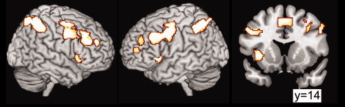
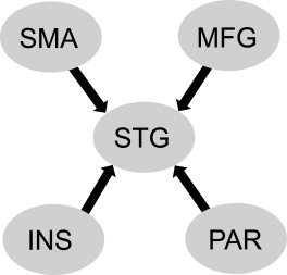
Methods: Subjects with an at risk mental state (ARMS) for psychosis, a first psychotic episode (FEP), and controls were studied using fMRI while performing a working memory task. Activation in the superior temporal gyrus (STG) was assessed using statistical parametric mapping, and its relationship to frontal activation was examined using dynamic causal modeling.
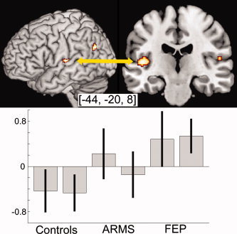
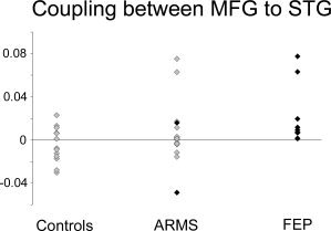
Results: The STG was differentially engaged across the three groups. There was deactivation of this region during the task in controls, whereas subjects with FEP showed activation and the response in subjects with ARMS was intermediately relative to the two other groups. There were corresponding differences in the effective connectivity between the STG and the middle frontal gyrus across the three groups, with a negative coupling between these areas in controls, a positive coupling in the FEP group, and an intermediate value in the ARMS group.
Conclusions: A failure to deactivate the superior temporal lobe during tasks that engage prefrontal cortex is evident at the onset of schizophrenia and may reflect a disruption of fronto-temporal connectivity. Qualitatively similar alterations are evident in people with prodromal symptoms of the disorder.
2009 Wiley-Liss, Inc.
Figures
References
-
- Allen P, Amaro E, Fu CH, Williams SC, Brammer MJ, Johns LC, McGuire PK ( 2007): Neural correlates of the misattribution of speech in schizophrenia. Br J Psychiatry 190: 162–169. - PubMed
-
- Borgwardt SJ, Riecher‐Rossler A, Dazzan P, Chitnis X, Aston J, Drewe M, Gschwandtner U, Haller S, Pfluger M, Rechsteiner E, D'souza M, Stieglitz RD, Radu EW, McGuire PK ( 2007): Regional gray matter volume abnormalities in the at risk mental state. Biol Psychiatry 61: 1148–1156. - PubMed
-
- Borgwardt SJ, McGuire PK, Aston J, Gschwandtner U, Pflüger MO, Stieglitz RD, Radue EW, Riecher‐Rössler A ( 2008): Reductions in frontal, temporal and parietal volume associated with the onset of psychosis. Schizophr Res 106( 2/3): 108–114. - PubMed
-
- Broome MR, Woolley JB, Johns LC, Valmaggia LR, Tabraham P, Gafoor R, Bramon E, McGuire PK ( 2005): Outreach and support in south London (OASIS): Implementation of a clinical service for prodromal psychosis and the at risk mental state. Eur Psychiatry 20( 5/6): 372–378. - PubMed
-
- Broome MR, Johns LC, Valli I, Woolley JB, Tabraham P, Brett C, Valmaggia L, Peters E, Garety PA, McGuire PK ( 2007): Delusion formation and reasoning biases in those at clinical high risk for psychosis. Br J Psychiatry 191 ( Suppl 51): s38–s42. - PubMed
Publication types
MeSH terms
Grants and funding
LinkOut - more resources
Full Text Sources
Medical
