

Interaction and conformational dynamics of membrane-spanning protein helices
- PMID: 19530249
- PMCID: PMC2775205
- DOI: 10.1002/pro.154
Interaction and conformational dynamics of membrane-spanning protein helices
Abstract
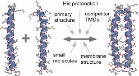
Within 1 or 2 decades, the reputation of membrane-spanning alpha-helices has changed dramatically. Once mostly regarded as dull membrane anchors, transmembrane domains are now recognized as major instigators of protein-protein interaction. These interactions may be of exquisite specificity in mediating assembly of stable membrane protein complexes from cognate subunits. Further, they can be reversible and regulatable by external factors to allow for dynamic changes of protein conformation in biological function. Finally, these helices are increasingly regarded as dynamic domains. These domains can move relative to each other in different functional protein conformations. In addition, small-scale backbone fluctuations may affect their function and their impact on surrounding lipid shells. Elucidating the ways by which these intricate structural features are encoded by the amino acid sequences will be a fascinating subject of research for years to come.
Figures


References
-
- Fleming KG. Riding the wave: structural and energetic principles of helical membrane proteins. Curr Opin Biotechnol. 2000;11:67–71. - PubMed
-
- Ubarretxena-Belandia I, Engelman DM. Helical membrane proteins: diversity of functions in the context of simple architecture. Curr Opin Struct Biol. 2001;11:370–376. - PubMed
-
- Arkin IT. Structural aspects of oligomerization taking place between the transmembrane alpha-helices of bitopic membrane proteins. Biochim Biophys Acta. 2002;1565:347–363. - PubMed
-
- Popot J-L, Engelman DM. Helical membrane protein folding, stability and evolution. Annu Rev Biochem. 2000;69:881–922. - PubMed
-
- Langosch D, Lindner E, Gurezka R. In vitro selection of self-interacting transmembrane segments—membrane proteins approached from a different perspective. IUBMB Life. 2002;54:1–5. - PubMed
Publication types
MeSH terms
Substances
LinkOut - more resources
Full Text Sources
