Review
. 2009 May;20(3):365-74.
doi: 10.1016/j.semcdb.2008.12.012.

Mitochondrial fusion and division: Regulation and role in cell viability

Affiliations
Review

Mitochondrial fusion and division: Regulation and role in cell viability

Giovanni Benard et al. Semin Cell Dev Biol. 2009 May.

Abstract

Discovery of various molecular components regulating dynamics and organization of the mitochondria in cells, together with novel insights into the role of mitochondrial fusion and division in the maintenance of cellular homeostasis, have provided some of the most exciting breakthroughs in the last decade of mitochondrial research. The focus of this review is on the regulation of mitochondrial fusion and division machineries. The newly identified factors associated with mitofusin/OPA1-dependent mitochondrial fusion, and Drp1-dependent mitochondrial division are discussed. Furthermore, the most recent findings on the role of mitochondrial fusion and division in the maintenance of cell function are also reviewed here in some detail.


Figures

Figure 1
Figure 1
Ultrastructure and cellular organization of mitochondria A) Mitochondrial structure in HeLa cell, as revealed by transmission electron microscopy (TEM). Black arrow shows the OMM, white arrow the IMM, and arrowheads show the IMM invaginations, called mitochondrial cristae. B) Network-like organization of mitochondria in cultured cells. HeLa cells were stained with anti-cytochrome c antibodies and imaged using confocal microscopy. C) Dynamic reorganization of mitochondria in cultured HeLa cells. Cells were transfected with photoactivable green fluorescent protein targeted to mitochondrial matrix (mito-PAGFP; for details see reference [28]), and then imaged with time-lapse confocal microscopy, after photoactivation of the small area of the cell. Note the diffusion of photoactivated mito-PAGFP (red rectangles), associated with remodeling of mitochondrial structure. These changes occur through balanced mitochondrial fusion and division processes.
Figure 2
Figure 2
Regulation of mitochondrial morphology by fusion and disvision A) An overview of proteins implicated in the regulation of mitochondrial fusion and division. For details see sections 3 and 4. B) Mitochondrial fusion and division regulate cellular organization of these organelles. Anti-cytochrome c antibody-immunostained mitochondria in Mfn2−/− MEFs (left) [12] and Mfn2−/− MEFs expressing a dominant negative mutant of MARCH5 are shown (for details see references [24,63,64]). These data indicate that mitochondrial fragmentation due to decline in fusion (left panel) can be reversed by relatively stronger defect in fission (right panel).
Figure 3
Figure 3
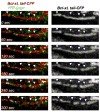
The large GTPase, Drp1 mediates division of mitochondria Drp1 is recruited to the mitochondria (arrowheads in the left panels) and this coincides and initiates mitochondrial division (arrowheads in the right panels) via the GTPase dependent mechanism (for details see references [23,25,27] and sections 2 and 4.2). HeLa cells expressing Drp1-YFP (green) and a marker of the OMM fused to CFP (Bcl-xL tail-CFP; red) were treated with Ca2+ ionophore, A23187, to initiate mitochondrial fission, followed by time-lapse confocal microscopy.

Similar articles

Cited by

References

    1. Brookes PS, Yoon Y, Robotham JL, Anders MW, Sheu SS. Calcium, ATP, and ROS: a mitochondrial love-hate triangle. Am J Physiol Cell Physiol. 2004;287:C817–833. - PubMed
    1. Wallace DC. A mitochondrial paradigm of metabolic and degenerative diseases, aging, and cancer: a dawn for evolutionary medicine. Annu Rev Genet. 2005;39:359–407. - PMC - PubMed
    1. Goyal G, Fell B, Sarin A, Youle RJ, Sriram V. Role of mitochondrial remodeling in programmed cell death in Drosophila melanogaster. Dev Cell. 2007;12:807–816. - PMC - PubMed
    1. Suen DF, Norris KL, Youle RJ. Mitochondrial dynamics and apoptosis. Genes Dev. 2008;22:1577–1590. - PMC - PubMed
    1. Jagasia R, Grote P, Westermann B, Conradt B. DRP-1-mediated mitochondrial fragmentation during EGL-1-induced cell death in C. elegans. Nature. 2005;433:754–760. - PubMed

Publication types

LinkOut - more resources