. 2009 Jun;16(6):822-32.
doi: 10.1016/j.devcel.2009.05.007.

Distinct origins and genetic programs of head muscle satellite cells

Affiliations

Distinct origins and genetic programs of head muscle satellite cells

Itamar Harel et al. Dev Cell. 2009 Jun.

Abstract

Adult skeletal muscle possesses a remarkable regenerative capacity, due to the presence of satellite cells, adult muscle stem cells. We used fate-mapping techniques in avian and mouse models to show that trunk (Pax3(+)) and cranial (MesP1(+)) skeletal muscle and satellite cells derive from separate genetic lineages. Similar lineage heterogeneity is seen within the head musculature and satellite cells, due to their shared, heterogenic embryonic origins. Lineage tracing experiments with Isl1Cre mice demonstrated the robust contribution of Isl1(+) cells to distinct jaw muscle-derived satellite cells. Transplantation of myofiber-associated, Isl1-derived satellite cells into damaged limb muscle contributed to muscle regeneration. In vitro experiments demonstrated the cardiogenic nature of cranial- but not trunk-derived satellite cells. Finally, overexpression of Isl1 in the branchiomeric muscles of chick embryos inhibited skeletal muscle differentiation in vitro and in vivo, suggesting that this gene plays a role in the specification of cardiovascular and skeletal muscle stem cell progenitors.
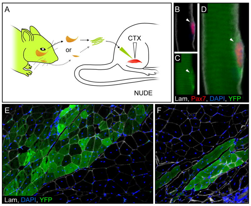

Figures

Figure 1
Figure 1. Head muscles and their associated satellite cells share a common mesoderm origin
A,B, Diagrams of the quail–chick small and large grafts (B) experiments suggesting that quail myoblasts contributed myonuclei and satellite cells in proportion to the graft size. C, A cartoon depicting selected muscle groups in an adult chick head. Green dots in the dorsal oblique (2) and mandibular adductors (3) represent quail nuclei. The dashed line indicates the plane of sectioning. D–F, Transverse sections stained for MyHC (red), quail-specific marker QCPN (green), and DAPI (blue) of eye and mastication muscles from post-hatch quail–chick chimera, using either large (D–E) or small (F) grafts. The dashed line indicates a region with high concentration of quail nuclei. G–I, Muscle sections (D–F) are followed by their respective higher magnifications stained with QPCN (peri-nuclear, green), Pax7 (nuclear, red), DAPI (blue) and laminin (Lam, white). Quail mesoderm-derived Pax7+ satellite cells located under the basal lamina are marked by full arrowhead; chick-derived satellite cells (empty arrowhead) can be seen in the small graft experiment (I). Scale bars, 150 μm (G), 5 μm (H–J).
Figure 2
Figure 2. Cranial (MesP1+) and trunk (Pax3+) derived muscles and satellite cells are genetically distinct
A, A cartoon of skeletal muscles derived from Mesp1Cre; RosaYFP crosses depicting representative trunk and head muscles, gastrocnemius (Gas) and masseter (Mas), respectively. B–I, Muscle and satellite cell analyses in a P10 Mesp1Cre; RosaYFP mouse reveal that the gastrocnemius is YFP (B) whereas the masseter is YFP+ (F). C–I, Immunofluorescence of transverse sections in these muscles showing both myofibers and associated satellite cells (arrowheads). J, A similar cartoon of Pax3Cre; RosaYFP crosses. K–R, Muscle and satellite cell analyses in P10 Pax3Cre; RosaYFP mice. The gastrocnemius muscle and associated satellite cells (arrowheads) are YFP+ (K,N) whereas the masseter is YFP- (O,R). Other Pax3 derived lineages such as the neural crest are revealed by the YFP+ staining in the facial nerve (FN). Scale bars: 5 μm.
Figure 3
Figure 3. Genetic heterogeneity of craniofacial muscles and satellite cells: Evidence of Isl1-derived satellite cells in branchiomeric muscles
A–E, Transverse sections of E16.5 mouse heads stained for MyHC (red), GFP/YFP (green), and DAPI (blue) of MesP1Cre (A), Pax3Cre (B), Isl1Cre (C) Nkx2.5Cre (D) and Myf5Cre (E) mice, crossed with Z/EG or RosaYFP (D) reporter lines. F, An anatomical cartoon of adult mouse head highlighting the lineage composition of distinct craniofacial muscle groups. G–V, Immunofluorescence of transverse sections in various cranial muscles in the Isl1Cre; RosaYFP (GR) and the masseter in the VE-CadCre; RosaYFP (S–V) mice. YFP+ cells are shown in green, Pax7+ (red), DAPI+ (blue) and laminin (Lam, white). Arrowheads and empty arrowheads indicate YFP+ and YFP satellite cells, respectively. S–V, Immunofluorescence for the VE-Cad (GFP) along with skeletal muscle (S, MyHC), satellite cells (T, Pax7 marked by empty arrowheads), smooth muscle (U, αSMA) and newly formed blood vessels (V, CD34). W, A table summarizing the percentage of YFP+ satellite cells in distinct YFP+ head muscles (green background). Scale bar: 5 μm.
Figure 4
Figure 4. Branchiomeric muscle-derived satellite cells can regenerate injured limb muscles
A, A diagram showing transplantation of single myofibers from the masseter and digastric of three week-old Myf5Cre; RosaYFP or Isl1Cre; RosaYFP mice, respectively, injected into the leg muscles of an age-matched, cardiotoxin-injected (CTX) immune-deficient (nude) mouse. B–D, Immunofluorescence of a sagital section in the masseter muscle from a Myf5Cre; RosaYFP mouse, revealing a YFP+ satellite cell (arrowhead, B–D) six-weeks post-transplantation. A transverse section in the transplanted gastrocnemius muscle revealed many YFP+ fibers originating from either the masseter of Myf5Cre; RosaYFP (E), or the digastric of Isl1Cre; RosaYFP (F).
Figure 5
Figure 5. Molecular analyses of cranial and trunk- derived satellite cell cultures
A, X-gal staining of satellite cell cultures derived from a Myf5-nLacZ mouse. B, RT-PCR and RT-qPCR analyses of satellite cells derived from selected cranial and trunk muscles, cultured with or without 200 ng/ml BMP4. C, A diagram summarizing shared and distinct effects of BMP4 on embryonic and adult, head and trunk mesoderm cells and satellite cells, respectively. gastrocnemius, (Gas); masseter (Mas); digastricus (Dig).
Figure 6
Figure 6. Isl1 represses head muscle differentiation in chick embryos, in vitro and in vivo
A, A diagram showing implantation of a bead soaked in 100 ng/ml BMP4, into the right CPM of a St.10 embryo. Whole-mount in situ hybridization for Isl1 (B–C) and the cross-section trough BA1 (D), 24h post-implantation, showing up-regulation of Isl1 in the implanted (right) BA1 (arrowheads) and down-regulation of Isl1 in the trigeminal ganglia (black arrowhead). An asterisk marks the bead in C. E, A diagram showing the CPM explant culture system. F, RT-PCR analysis of CPM explants after 0–4 days in culture. G, RCAS-Isl1 but not RCAS-GFP overexpression resulted in inhibition of myogenesis in CPM explants. H, A diagram showing retroviral injection, at the CPM of a St. 8 chick embryo, developed until E5 for further analysis. J–O, Immunofluorescence of transverse sections through BA1 in an E5 chick embryo, infected unilaterally with either control RCAS-GFP (I–K) or RCAS-Isl1 (L–N). Arrows indicate differentiating myotubes, which were partially repressed by Isl1 (empty arrow). TG, trigeminal ganglia. BA, branchial arch.
Figure 7
Figure 7. Distinct myogenic programs in the embryo contribute to skeletal muscles and satellite cells in the head and trunk
A. Satellite cells, located on the surface of the myofiber, beneath its basement membrane (basal lamina), serve as a source of myogenic cells for growth and repair of postnatal skeletal muscle. B. The combinatorial use of several mouse Cre lines, depicted as circles, allowed us to draw sharp boundaries between head muscle groups (tongue, mastication, eye muscles) as marked by the overlap between the lineages. The contribution of these mouse Cre lines to other mesoderm lineages is depicted in brackets. C. Muscles and satellite cells in trunk and limb derive from somites (Pax3 lineage) while branchiomeric muscles (including the mastication muscles) and their associated satellite cells derive from both CPM and SpM sources (MesP1, Isl1, and Nkx2.5 lineages). Our findings reveal the lineage signature of the head musculature and its associated satellite cell population, highlighting the overwhelming heterogeneity in these groups of muscles.

Comment in

References

    1. Alva JA, Zovein AC, Monvoisin A, Murphy T, Salazar A, Harvey NL, Carmeliet P, Iruela-Arispe ML. VE-Cadherin-Cre-recombinase transgenic mouse: a tool for lineage analysis and gene deletion in endothelial cells. Dev Dyn. 2006;235:759–767. - PubMed
    1. Armand O, Boutineau AM, Mauger A, Pautou MP, Kieny M. Origin of satellite cells in avian skeletal muscles. Arch Anat Microsc Morphol Exp. 1983;72:163–181. - PubMed
    1. Asakura A, Komaki M, Rudnicki M. Muscle satellite cells are multipotential stem cells that exhibit myogenic, osteogenic, and adipogenic differentiation. Differentiation. 2001;68:245–253. - PubMed
    1. Beauchamp JR, Heslop L, Yu DS, Tajbakhsh S, Kelly RG, Wernig A, Buckingham ME, Partridge TA, Zammit PS. Expression of CD34 and Myf5 defines the majority of quiescent adult skeletal muscle satellite cells. J Cell Biol. 2000;151:1221–1234. - PMC - PubMed
    1. Bothe I, Ahmed MU, Winterbottom FL, von Scheven G, Dietrich S. Extrinsic versus intrinsic cues in avian paraxial mesoderm patterning and differentiation. Dev Dyn. 2007;236:2397–2409. - PubMed

Publication types

MeSH terms

Substances