. 2009 Jun;16(6):844-55.
doi: 10.1016/j.devcel.2009.05.013.

Effects of age on meiosis in budding yeast

Affiliations

Effects of age on meiosis in budding yeast

Monica Boselli et al. Dev Cell. 2009 Jun.

Abstract

In humans, the frequency with which meiotic chromosome mis-segregation occurs increases with age. Whether age-dependent meiotic defects occur in other organisms is unknown. Here, we examine the effects of replicative aging on meiosis in budding yeast. We find that aged mother cells show a decreased ability to initiate the meiotic program and fail to express the meiotic inducer IME1. The few aged mother cells that do enter meiosis complete this developmental program but exhibit defects in meiotic chromosome segregation and spore formation. Furthermore, we find that mutations that extend replicative life span also extend the sexual reproductive life span. Our results indicate that in budding yeast, the ability to initiate and complete the meiotic program as well as the fidelity of meiotic chromosome segregation decrease with cellular age and are controlled by the same pathways that govern aging of asexually reproducing yeast cells.


Figures
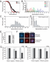
Figure 1
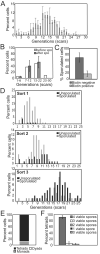
Figure 1. A method to study the effects of replicative age on meiosis in yeast
(A) The life-span of wild-type W303 (A702, closed squares) and wild-type SK1 (A727, open squares) cells was compared by single-cell analysis. The experiment was performed in triplicate using at least 40 cells for each experiment. Each cell that we analyzed was the granddaughter of the cell initially isolated at random from the population. The median life-span for W303 was 19.7 (SD=0.58) and the mean life-span 18.9 (SD=0.96). For SK1 the median life-span was 21.5 (SD=2.29) and mean life-span 22.5 (SD=0.95). (B) The age distribution of biotin positive cells (A16554) after each sort was determined by counting the number of bud scars (n>50 for each experiment). (C) The age distribution of biotin positive cells (A16554) after the third sort was determined by counting the number of bud scars (n>50 for each experiment). (D) The age distribution of biotin negative cells (A16554) after the third sort was determined by counting the number of bud scars (n>50 for each experiment). (E, F) The percentage of cells (A16554) with a fragmented nucleolus as judged by Nop1 staining was determined in biotin positive cells obtained from sort 3 (E; n=200 for biotin negative cells; n=400 for biotin positive cells). The picture in (F) shows a cell with an intact nucleolus (top) and a cell with a fragmented nuclelus (bottom). Nop1 is shown in red, DNA in blue. (G, H) After each sort, biotin positive and biotin negative cells (A16554) were plated on YEP+ 2% glucose plates (E) or YEP + 2% glycerol plates (F) and the number of colonies that formed were counted.
Figure 2
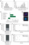
Figure 2. Aged cells fail to sporulate
(A–C) Cells were grown and sorted as described in Experimental Procedures to obtain biotin positive old cells and biotin negative new-born cells. Biotin positive cells (A16554) with the age distribution shown in (A) were induced to sporulate and the age distribution in cultures was determined before and after incubation in sporulation medium (B). The sporulation efficiency was determined 24 hours after transfer in sporulation medium (N=8 experiments) (C). (D) Cells (A16554) were grown and sorted as described in Experimental Procedures. After sort 1 a fraction of biotin positive cells was induced to sporulate (Sort1). After 24 hours the number of bud scars per cell was determined and correlated with the cell's ability to sporulate. The cells obtained from sort 2 and sort 3 were analyzed in the same manner (n=100). The probability of results from different sorts being the same is as follows: Sort 1 and sort 2 p=0.0001096, sort1 and sort 3 p=1,6×10⁻²⁰, sort 2 and sort 3 p=2,3×10⁻⁸ . (E, F) Biotin negative cells (A16554) were induced to sporulate. After 24 hours sporulation efficiency (C) and the percentage of monads, dyads and tetrads (D) was determined. The number of viable spores in tetrads is shown in (E) (n=240 tetrads).
Figure 3
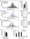
Figure 3. Aged cells do not enter the meiotic program
(A–C) Wild-type cells (A16554) were grown and sorted as described in Experimental Procedures. Cells sorted either once (young cells, age distribution shown in (A)) or three times (old cells; age distribution shown in (B)) were induced to sporulate for 6 hrs to determine their gene expression pattern. (C) shows average changes in transcript levels for genes identified as up-regulated during meiosis in Primig et al. (2000). Grouped clusters identified by Primig et al. are shown on the y axis. Abbreviations: DS = DNA synthesis, Rec = Recombination, SC = sister chromatid cohesion, preM = pre-meiotic, MI = Meiosis I, M2 = meiosis II. (D – H) Cells carrying the NLS-mCherry fusion under the control of the IME1 promoter (A18489) were grown and sorted as described in Experimental Procedures and induced to sporulate in standard sporulation medium. The presence of mCherry was analyzed at the indicated times in biotin positive cells with the age distribution shown in (E) and biotin negative cells (F). (D) shows an old cell that fails to express IME1-NLS-mCHERRY and a new-born cell that does. Bud scars are shown in blue, IME1-NLS-mCherry fusion in red. (G, H) Cells carrying the IME1-NLS-mCHERRY fusion (A18489) were grown and sorted as described in Experimental Procedures. Then the biotin positive (median age= 16 generations; age distribution in G) and biotin negative populations were combined and induced to enter meiosis (H). The ability to express mCherry was determined for each population at the indicated times (n>100).
Figure 4
Figure 4. Overexpression of IME1 partially suppresses the sporulation defect of aged cells
(A-C) Cells carrying IME1 under the control of the CUP1 promoter (pCUP1-IME1; A22260) were grown in YPA for 12 hours, washed and then transferred to sporulation medium (0.3 % KOAc, 0.02% raffinose) containing 50μM of CuSO. ~8 χ 107 cells were used for protein extraction (TCA) for each time point. (B, C) Cells heterozygous for the pCUP1-IME1 fusion (A22764) were grown as described in Experimental Procedures to obtain biotin positive old cells (mean age = 15.73 generations, SD= 4.96) with the age distributions shown in (B) were either transferred to sporulation-inducing conditions either in the presence (+CuSO4) or absence (−CuSO4) of CuSO4 . The percentage of sporulated cells was determined after 24 hours. P=1.025×10⁻⁹ .
Figure 5
Figure 5. Increased meiosis II non-disjunction in aged cells
(A–D) Cells carrying GFP dots at LYS2 on both copies of chromosome II (homozygous LYS2-GFP dots; A16554) were grown and sorted as described in Experimental Procedures. Biotin positive cells with the age distribution shown in (A) and biotin negative cells were induced to sporulate in preconditioned medium. After 24 hours overall sporulation efficiency (B; n>100 cells) and the number of bud scars per cell was determined and correlated with the cell's ability to sporulate (C; n=95). Unsporulated cells are shown in grey, sporulated cells with a wild-type distribution of GFP dots are shown in blue and cells with GFP dots in only three of the four nuclei are shown in red. The overall GFP dot distribution in biotin positive and biotin negative cells during both Meiosis I and Meiosis II are shown in (D; n=100/experiment; binucleate analysis top panel; tetranucleate analysis bottom panel). Note that in 43% of aged cells had a daughter cell attached to it. In the event that this daughter cell sporulated (40%) the GFP dot segregation pattern was not examined. (E) Aged mother cells (A16554) were isolated and induced to sporulate in preconditioned medium. The ability of cells to package the four meiotic products into spores was correlated with the number of bud scars. The results represent data from three independent experiments (n=112). (F, G) Tetrads obtained from aged mother cells (biotin positive cells; average age =15.72 SD= 5.53; A16554) were identified and dissected using a fluorescence/dissecting microscope. Percent of viable spores (N= 200 spores) is shown in (F) and the number of viable spores per tetrad (N=50 tetrads) in (G).
Figure 6
Figure 6. A mutation that extends the replicative life span suppresses the sporulation defect of aged cells
(A–D) Wild type cells (A16554) and cells lacking FOB1 (A18319) were grown and sorted as described in Experimental Procedures. Biotin positive cells with the age distribution shown in (A) and biotin negative cells were induced to sporulate. After 24 hours the number of bud scars per cell was determined and correlated with the cell's ability to sporulate (B; n=100). Tetrads obtained from fob1Δcells were dissected to determine spore viability (C; n=160) and the number of viable spores in tetrads (D, n=40 tetrads). (E, F) Cells overexpressing SIR2 (A20375) were grown and sorted as described in Experimental Procedures. Biotin positive cells with the age distribution shown in (E) and biotin negative cells were induced to sporulate for 24 hours to determine the number of sporulated cells (F; n>100). (G, H) Wild type (A16554; G) and sir2Δ(A20782; H) cells were sorted one time by FACS and induced to sporulate. Biotin positive (median age 6 generations) and biotin negative cells (median age 2 generations) were sporulated for 24 hours to determine the number of sporulated cells (n>100).
Figure 7
Figure 7. ERCsdecrease the ability of the cells to enter meiosis
(A, B) Biotin positive cells harboring a 2μ plasmid carrying the FOB1 gene (A22532) were first grown in medium lacking uracil, labeled with biotin, then grown in presporulation medium (YEP+2% potassium acetate; YPA) for few generations and sorted one time to obtain young and new-born cells. Biotin positive young cells with the age distribution shown in (A) and biotin negative new-born cells were induced to sporulate for 24 hours to determine the number of sporulated cells (n>100) (B). (C, D) Cells harboring a 2μ plasmid carrying the 5S and the 35S rDNA repeats (A22611) were first grown in medium lacking leucine, labeled with biotin, then grown in presporulation medium (YEP+2% potassium acetate; YPA) for few generations and sorted one time to obtain young and new-born cells. Biotin positive young cells with the age distribution shown in (C) and biotin negative new-born cells were induced to sporulate for 24 hours to determine the number of sporulated cells (n>100) (B).

References

    1. Bonawitz ND, Rodeheffer MS, Shadel GS. Defective mitochondrial gene expression results in reactive oxygen species-mediated inhibition of respiration and reduction of yeast life span. Mol Cell Biol. 2006;26:4818–29. - PMC - PubMed
    1. Colomina N, Liu Y, Aldea M, Garí E. TOR regulates the subcellular localization of Ime1, a transcriptional activator of meiotic development in budding yeast. Mol Cell Biol. 2003;23:7415–24. - PMC - PubMed
    1. Covitz PA, Herskowitz I, Mitchell AP. The yeast RME1 gene encodes a putative zinc finger protein that is directly repressed by a1-alpha 2. Genes Dev. 1991;5:1982–9. - PubMed
    1. Chen C, Dewaele S, Braeckman B, Desmyter L, Verstraelen J, Borgonieb G, Vanfleterenb J, Contrerasa R. A high-throughput screening system for genes extending life-span. Experimental Gerontology. 2003;38:1051–1063. - PubMed
    1. Defossez PA, Prusty R, Kaeberlein M, Lin SJ, Ferrigno P, Silver PA, Keil RL, Guarente L. Elimination of the replication block protein Fob1 extends the life span of yeast mother cells. Mol. Cell. 1999;3:447–55. - PubMed

Publication types

MeSH terms

Associated data