. 2009 Aug 14;284(33):22022-22028.
doi: 10.1074/jbc.M109.032235. Epub 2009 Jun 15.

Cytotoxicity mediated by the Fas ligand (FasL)-activated apoptotic pathway in stem cells

Affiliations

Cytotoxicity mediated by the Fas ligand (FasL)-activated apoptotic pathway in stem cells

Julia Mazar et al. J Biol Chem. .

Abstract

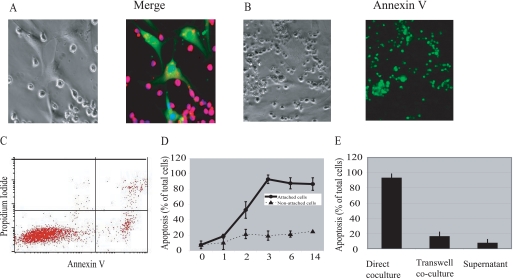
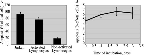
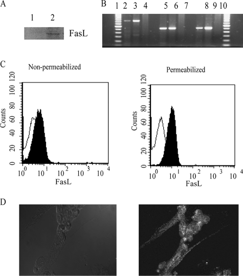
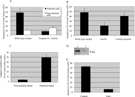
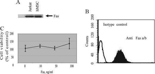
Whereas it is now clear that human bone marrow stromal cells (BMSCs) can be immunosuppressive and escape cytotoxic lymphocytes (CTLs) in vitro and in vivo, the mechanisms of this phenomenon remain controversial. Here, we test the hypothesis that BMSCs suppress immune responses by Fas-mediated apoptosis of activated lymphocytes and find both Fas and FasL expression by primary BMSCs. Jurkat cells or activated lymphocytes were each killed by BMSCs after 72 h of co-incubation. In comparison, the cytotoxic effect of BMSCs on non-activated lymphocytes and on caspase-8(-/-) Jurkat cells was extremely low. Fas/Fc fusion protein strongly inhibited BMSC-induced lymphocyte apoptosis. Although we detected a high level of Fas expression in BMSCs, stimulation of Fas with anti-Fas antibody did not result in the expected BMSC apoptosis, regardless of concentration, suggesting a disruption of the Fas activation pathway. Thus BMSCs may have an endogenous mechanism to evade Fas-mediated apoptosis. Cumulatively, these data provide a parallel between adult stem/progenitor cells and cancer cells, consistent with the idea that stem/progenitor cells can use FasL to prevent lymphocyte attack by inducing lymphocyte apoptosis during the regeneration of injured tissues.


Figures

FIGURE 1.
FIGURE 1.
Lymphocyte apoptosis induced by BMSCs. A, Jurkat cells and BMSCs were labeled with CellTracker Red and Green, respectively. Jurkat cells were incubated with a monolayer of BMSCs. Two hours following incubation, the samples were stained with Hoechst, a nuclear dye, and analyzed by confocal microscopy. BMSCs induce apoptosis in lymphocytes through cell-cell contact. Jurkat cells were added to the monolayer of BMSCs in a ratio of 1:10 directly (B–D) or separated by the transwell inserts, or supernatant from BMSCs-Jurkat co-culture was collected 18 h after co-culture initiation and added to control Jurkat cells (E). After the indicated time periods, non-attached Jurkat cells were removed from the co-culture chambers. The percentage of annexin V-positive cells among attached cells was detected by fluorescent microscopy (B) and among non-attached cells, by flow cytometry (C). The results of three independent experiments have been summarized ± S.E. (D and E).
FIGURE 2.
FIGURE 2.
Apoptosis is specific for BMSCs and activated lymphocytes co-culture. PHA-activated and non-activated lymphocytes were added to monolayer of BMSCs. Non-attached lymphocytes were removed after 24 h of co-incubation. The percentage of annexin V-positive attached lymphocytes was detected by fluorescent microscopy (A). Jurkat cells were added to the monolayer of skin fibroblasts in a ratio of 1:10. After the indicated time periods, non-attached Jurkat cells were removed from the co-culture chambers. The percentage of annexin V-positive cells among attached cells was detected by fluorescent microscopy (B). The results of three independent experiments are presented as mean ± S.E.
FIGURE 3.
FIGURE 3.
Expression of FasL by BMSCs. A, immunoblot detection of FasL in BMSC lysates. Expression of FasL was assessed by Western blot analysis using a polyclonal rabbit anti-human FasL antibody. Cells lysates were prepared separately from BMSCs and lymphocytes, not under co-culture conditions. B, RT-PCR analysis of human Fas ligand in BMSCs. Lanes 1 and 11 correspond to the 100 bp DNA ladder from Invitrogen. Lanes 2–4 correspond to almost the full-length Fas ligand (845 base pairs) with primers FASL5 and FASL8. Lanes 5–7 correspond to the first half of the full-length Fas ligand (518 base pairs) with primers FASL 5 and FASL 6. Lanes 8–10 correspond to the second half of the full-length Fas ligand (516 base pairs) with primers FASL 7 and FASL 8. C, flow cytometry analysis of FasL expression on permeabilized and non-permeabilized BMSCs using fluorescein isothiocyanate-conjugated monoclonal anti-FasL antibody. D, fluorescent microscopy analysis of FasL expression on BMSC following 24 h co-culture with Jurkat cells.
FIGURE 4.
FIGURE 4.
Role of death receptors in Jurkat apoptosis initiated in co-culture with BMSCs. Wild type (WT) and caspase-8-negative Jurkat cells were incubated with a monolayer of BMSCs at a ratio of 1:100 for 48 h (A). Four hours after initiation of co-culture, non-attached Jurkat cells were removed from the co-culture chambers. At the end of co-incubation, all attached cells were trypsinized and labeled with annexin V and anti-CD3 antibody. The percentage of annexin V-positive cells among CD3-positive, attached cells was detected by flow cytometry. Separately, Jurkat cells were co-incubated with BMSCs for 48 h, non-attached cells were removed, and annexin V labeling of non-attached cells was detected by flow cytometry. Fas Fc fusion protein (1 μg/ml) and control Fc protein (1 μg/ml) were added to BMSCs 10 min prior to co-culture initiation (B). Caspase-3 activity of attached Jurkat cells was evaluated using the fluorescent substrate for caspase-3 24 h after co-culture initiation (C). BMSCs were transiently transfected with FasL siRNA or negative control siRNA. Down-regulation of FasL expression by specific siRNA was detected by Western blot (D). Jurkat cells were incubated with a monolayer of wild type and FasL siRNA-transfected BMSCs at a ratio of 1:100 for 48 h. Four hours after initiation of co-culture, the percentage of annexin V-positive cells among CD3-positive, attached cells was detected by microscopy (E).
FIGURE 5.
FIGURE 5.
Expression and functionality of Fas on BMSCs. A, immunoblot detection of Fas in BMSC lysates. Expression of FasL was assessed by Western blot analysis using a monoclonal anti-human FasL antibody. B, flow cytometric analysis of surface Fas expression on BMSC using PE-conjugated monoclonal anti-human Fas antibody. C, BMSCs were incubated with different concentrations of CH-11 anti-Fas antibody for 18 h. Cell viability was assessed by the WST-1 dye assay.

References

    1. Robey P. G., Bianco P. (2006) J. Am. Dent. Assoc. 137, 961–972 - PubMed
    1. Friedenstein A. J., Piatetzky-Shapiro , II, Petrakova K. V. (1966) J. Embryol. Exp. Morphol. 16, 381–390 - PubMed
    1. Kemp K. C., Hows J., Donaldson C. (2005) Leuk. Lymphoma 46, 1531–1544 - PubMed
    1. Ferrari G., Cusella-De Angelis G., Coletta M., Paolucci E., Stornaiuolo A., Cossu G., Mavilio F. (1998) Science 279, 1528–1530 - PubMed
    1. Prockop D. J. (1997) Science 276, 71–74 - PubMed

Publication types