. 2009 Jun;7(6):841-50.
doi: 10.1158/1541-7786.MCR-08-0555. Epub 2009 Jun 16.

Zebrafish Hagoromo mutants up-regulate fgf8 postembryonically and develop neuroblastoma

Affiliations

Zebrafish Hagoromo mutants up-regulate fgf8 postembryonically and develop neuroblastoma

Adam Amsterdam et al. Mol Cancer Res. 2009 Jun.

Abstract

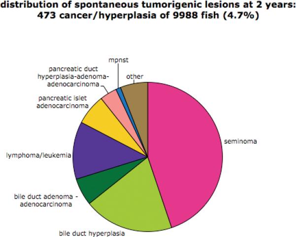
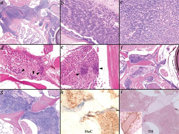
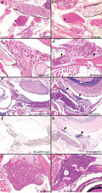
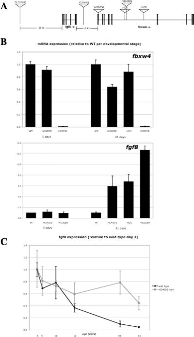
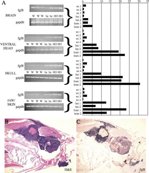
We screened an existing collection of zebrafish insertional mutants for cancer susceptibility by histologic examination of heterozygotes at 2 years of age. As most mutants had no altered cancer predisposition, this provided the first comprehensive description of spontaneous tumor spectrum and frequency in adult zebrafish. Moreover, the screen identified four lines, each carrying a different dominant mutant allele of Hagoromo previously linked to adult pigmentation defects, which develop tumors with high penetrance and that histologically resemble neuroblastoma. These tumors are clearly neural in origin, although they do not express catecholaminergic neuronal markers characteristic of human neuroblastoma. The zebrafish tumors result from inappropriate maintenance of a cell population within the cranial ganglia that are likely neural precursors. These neoplasias typically remain small but they can become highly aggressive, initially traveling along cranial nerves, and ultimately filling the head. The developmental origin of these tumors is highly reminiscent of human neuroblastoma. The four mutant Hagoromo alleles all contain viral insertions in the fbxw4 gene, which encodes an F-box WD40 domain-containing protein. However, although one allele clearly reduced the levels of fbxw4 mRNA, the other three insertions had no detectable effect on fbw4 expression. Instead, we showed that all four mutations result in the postembryonic up-regulation of the neighboring gene, fibroblast growth factor 8 (fgf8). Moreover, fgf8 is highly expressed in the tumorigenic lesions. Although fgf8 overexpression is known to be associated with breast and prostate cancer in mammals, this study provides the first evidence that fgf8 misregulation can lead to neural tumors.


Figures

Figure 1
Figure 1
Distibution of types of tumorigenic lesions in the background population.
Figure 2
Figure 2
Neuroblastoma-like tumors in Hag heterozygotes at two years of age. A. (40X) a very advanced tumor fills most of the head space between the esophagus and the skull and invades the body wall musculature; B-C. (400X) rosette arrangement of cells is occasionally observed in tumors (B) but more commonly cellular arrangement is more variable (C); D-E. (400X) very small neoplasias (black arrowheads) observed in the ganglia within the skull below the midbrain (D) or posterior of the ear (E); F. (100X) this tumor can be seen growing along the nerve running behind the eye as well as another below the skull; G-I. (200X) H&E (G), anti-HuC (H), and anti-TH (I) of the same tumor; inset in I shows TH staining of adrenal cells in the kidney on the same slide as a positive control for the antibody.
Figure 3
Figure 3
Tumors begin as inappropriate maintenance of a normal developmental stage of putative neural precursors. A-B. (400X) at 4 weeks small cells resembling the tumor cells (asterisks) are seen in cranial ganglia of both wild type (A) and Hag (B) fish; C-D. (400X) at 12 weeks, only fully developed ganglion cells are observed in wild type fish (C), but the small precursor-like cells (black arrowheads) are still observed in most Hag mutants (D). E-H. (200X) the small cells seen uniquely in Hag mutants (black arrowheads) at 12 weeks (F) stain strongly for HuC mRNA by in situ hybridization (H); ganglion cells in wild type (E) and Hag mutants (F) stain weakly for HuC (G,H); I. (400X) tumor beginning to spread over entire ganglia in 24 week Hag heterozygote; J. (100X) tumor in 31 week homozygote growing within the skull and along nerves behind and above the eye.
Figure 4
Figure 4
Hag mutations are insertions in the fbxw4 gene but affect expression of the neighboring fgf8 gene. A. Location of the mutagenic insertions in Hag mutants. hiD2058 is precisely at the splice donor of exon 1 of fbxw4; the other three insertions are in the 24 kb fifth intron. Enhancer trap insertions CLGY1030 and CLGY508 are in intergenic sequence upstream and downstream of fgf8, respectively. B. fbxw4 and fgf8 mRNA measured in whole fish by real-time RT-PCR in wild type and homozygotes of multiple Hag alleles at 5 and 41 days. Units are normalized to wild type sample of each developmental age. C. fgf8 mRNA measured in whole fish by real-time RT-PCR in wild type and hiD4000 homozygotes from 3 to 41 days. Units are normalized to the 3 day wild type sample. For B and C, error bars show the standard deviation of 2−3 technical replicates.
Figure 5
Figure 5
Hag mutants overexpress fgf8 in the head region of young adults and in tumors A. fgf8 mRNA in multiple parts of the head of 3 month old wild type, hiD2058 heterozygote and hiD2058 homozygote fish were measured by real-time and semi-quantitative RT-PCR; gapdh was used as a normalization control. Samples were isolated from three (brain) or four wild type fish and two each Haghi2058 heterozygotes and homozygotes. Ethidium bromide stained gels from semi-quantitative RT-PCR are shown on the left; quantification of the real-time PCR is shown on the right. Quantification is normalized within the dataset for each tissue to the average wild type sample of that tissue; error bars show the standard deviation of three technical replicates. B-C. (20X) H&E (B) and fgf8 in situ hybridization (C) on a very advanced tumor in a 2 year old hiD2058 homozygote.

References

    1. Feitsma H, Cuppen E. Zebrafish as a Cancer Model. Mol Cancer Res. 2008;6:685–94. - PubMed
    1. Amsterdam A, Sadler KC, Lai K, et al. Many ribosomal protein genes are cancer genes in zebrafish. PLoS Biol. 2004;2:E139. - PMC - PubMed
    1. Berghmans S, et al. tp53 mutant zebrafish develop malignant peripheral nerve sheath tumors. Proc Natl Acad Sci U S A. 2005;102:407–12. - PMC - PubMed
    1. Haramis AP, Hurlstone A, van der Velden Y, et al. Adenomatous polyposis coli-deficient zebrafish are susceptible to digestive tract neoplasia. EMBO Rep. 2006;7:444–9. - PMC - PubMed
    1. Feitsma H, Kuiper RV, Korving J, Nijman IJ, Cuppen E. Zebrafish with mutations in mismatch repair genes develop neurofibromas and other tumors. Cancer Res. 2008;68:5059–66. - PubMed

Publication types