

Review
doi: 10.1258/jrsm.2009.080240.
The expanding role of interventional radiology in head and neck surgery
Affiliations
- PMID: 19531617
- PMCID: PMC2697043
- DOI: 10.1258/jrsm.2009.080240
Item in Clipboard
Review
The expanding role of interventional radiology in head and neck surgery
J R Soc Med.
2009 Jun.
No abstract available
Figures

(a) Digital subtraction angiogram showing nasal vasculature with bleeding point (black arrow) and catheter (white arrow); (b) after selective embolization there is no blood flow to the bleeding point (black arrow). Blood flow to the anterior part of the nose has been preserved (white arrow)

(a) Digital subtraction angiogram showing nasal vasculature with bleeding point (black arrow) and catheter (white arrow); (b) after selective embolization there is no blood flow to the bleeding point (black arrow). Blood flow to the anterior part of the nose has been preserved (white arrow)
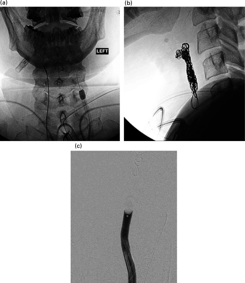
(a) Temporary balloon occlusion test. The inflated balloon is seen in the left common carotid artery of a patient with uncontrolled haemorrhage secondary to malignant erosion of the carotid artery; (b) lateral view of the same patient after coil embolization; (c) this patient required permanent balloon occlusion to fully control the haemorrhage

(a) DSA of juvenile nasal angiofibroma (JNA) showing microcatheter (black arrow) and tumour blush (white arrow); (b) after embolization there is minimal vascularity of the JNA

(a) DSA of juvenile nasal angiofibroma (JNA) showing microcatheter (black arrow) and tumour blush (white arrow); (b) after embolization there is minimal vascularity of the JNA

(a) Tumour blush of a large glomus tumour before embolization; (b) after embolization of the ascending pharyngeal artery with coils and particles the vascularity is reduced
References
-
- Rösch J, Keller FS, Kaufman JA. The birth, early years, and future of interventional radiology. J Vasc Interv Radiol 2003;14:841–53 - PubMed
-
- Becker GJ. Interventional radiology 2000 and beyond: back from the brink. J Vasc Interv Radiol 1999;10:681–7 - PubMed
-
- Keller FS. Interventional radiology: new paradigms for the new millennium. J Vasc Interv Radiol 2000;11:678–81 - PubMed
-
- Klotz DA, Winkle MR, Richmon J, Hengerer AS. Surgical Management of posterior epistaxis: a changing paradigm. Laryngoscope 2002;112:1577–82 - PubMed
-
- Snyderman CH, Goldman SA, Carrau RL, Ferguson BJ, Grandis JR. Endoscopic sphenopalatine artery ligation is an effective method of treatment for posterior epistaxis. Am J Rhinol 1999;13:137–40 - PubMed
Publication types
MeSH terms
LinkOut - more resources
Full Text Sources
Medical
