. 2009 Aug;37(15):5019-31.
doi: 10.1093/nar/gkp515. Epub 2009 Jun 16.

The Myb/SANT domain of the telomere-binding protein TRF2 alters chromatin structure

Affiliations

The Myb/SANT domain of the telomere-binding protein TRF2 alters chromatin structure

Asmaa M Baker et al. Nucleic Acids Res. 2009 Aug.

Abstract

Eukaryotic DNA is packaged into chromatin, which regulates genome activities such as telomere maintenance. This study focuses on the interactions of a myb/SANT DNA-binding domain from the telomere-binding protein, TRF2, with reconstituted telomeric nucleosomal array fibers. Biophysical characteristics of the factor-bound nucleosomal arrays were determined by analytical agarose gel electrophoresis (AAGE) and single molecules were visualized by atomic force microscopy (AFM). The TRF2 DNA-binding domain (TRF2 DBD) neutralized more negative charge on the surface of nucleosomal arrays than histone-free DNA. Binding of TRF2 DBD at lower concentrations increased the radius and conformational flexibility, suggesting a distortion of the fiber structure. Additional loading of TRF2 DBD onto the nucleosomal arrays reduced the flexibility and strongly blocked access of micrococcal nuclease as contour lengths shortened, consistent with formation of a unique, more compact higher-order structure. Mirroring the structural results, TRF2 DBD stimulated a strand invasion-like reaction, associated with telomeric t-loops, at lower concentrations while inhibiting the reaction at higher concentrations. Full-length TRF2 was even more effective at stimulating this reaction. The TRF2 DBD had less effect on histone-free DNA structure and did not stimulate the t-loop reaction with this substrate, highlighting the influence of chromatin structure on the activities of DNA-binding proteins.


Figures

Figure 1.
Figure 1.
Analysis of nucleosomal array fibers. The pRST5 plasmid and expected fragments created by PvuII or SfaNI digestion (A). Micrococcal nuclease digestion at indicated time points of nucleosomal arrays reconstituted onto PvuII (B); SfaNI (C) digested pRST5 DNA. Multi-gels of telomeric nucleosomal array fibers (NA) and histone-free DNA (DNA) from pRST5 digested with PvuII (D); and SfaNI (E) prepared and subjected to electrophoresis according to ‘Materials and Methods’ section. Spheres refer to carboxylate-coated microsphere standards (35 nm radius). The ‘1 kb tel’ and ‘2 kb tel’ refer to the telomeric fragments liberated by PvuII and SfaNI digestion respectively. ‘N’ refers to the fragments without telomeric DNA. Logarithmic plot of pore sizes (Pe) versus agarose% (F). Data was obtained from multi-gels run with bacteriophage T3 (Phage) in this laboratory or previous work [Phage, previous, (24)] and 35 nm carboxylate-coated microspheres (Microspheres). Pe for each agarose concentration was calculated according to ‘Materials and Methods’. Symbols with error bars represent the mean ± 1 SD of four to eight determinations.
Figure 2.
Figure 2.
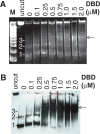
TRF2 DBD binds specifically to telomeric DNA and nucleosomal arrays. 0.6% agarose gels of TRF2 DBD binding to DNA (DNA) (A) and nucleosomal arrays (NA) (B) from the pRST5 fragment digested to obtain a 1-kb DNA fragment with 580-bp telomeric DNA (telo) and 2.5-kb non-telomeric DNA (N-telo). Gels similar to (A) and (B), respectively except the pRST5 was digested to obtain a 2-kb fragment containing the 580-bp telomeric DNA (telo) with a 1 kb and smaller fragments being non-telomeric (N-telo) (C and D).
Figure 3.
Figure 3.
TRF2 DBD-dependent changes in surface charge density (μ'0), effective radius (Re from dilute gels) and conformation flexibility (Re versus Pe). The μ'0 at each indicated TRF2 DBD concentration was obtained by Ferguson plots of mobilities derived from multi-gels with agarose concentrations of 0.25–1% according to ‘Materials and Methods’ section (A). The Re at each TRF2 DBD concentration derived from 0.25–1% multi-gel experiments according to ‘Materials and Methods’ section (B). The Re values from 0.25–0.6% agarose (dilute gels) were averaged from each multi-gel experiment. The μ'0 or Re for each TRF2 DBD concentration was normalized to 0 nM TRF2 DBD. Each data point represents the mean ± 1 SD of 3-7 multi-gel experiments. The effect of the TRF2 DBD on the reptation of DNA and nucleosomal arrays (change in Re versus Pe) in concentrated gels (C and D). Plots of effective radius (Re) versus pore size (Pe) of DNA (C) or nucleosomal arrays (D) incubated with indicated amounts of TRF2 DBD. Re and Pe were derived from multi-gels of 0.7–2.5% agarose concentrations or 0.4–2.1% agarose concentrations according to ‘Materials and Methods’ section. The DNA fragment analyzed in all experiments in this figure was the 2-kb telomeric fragment unless otherwise indicated.
Figure 4.
Figure 4.
TRF2 DBD-dependent changes in micrococcal nuclease digestion. Nucleosomal arrays (12.5 ng/ul) were incubated with indicated amounts of TRF2 DBD and digested with micrococcal nuclease according to ‘Materials and Methods’ section. Deproteinated samples were electrophoresed on a 12% native polyacrylamide gel and stained with SYBR Green (A) followed by transfer to a membrane and blotting with a biotin-d(TTAGGG)7 probe (B) as described in the ‘Material and methods’ section. M refers to 100-bp ladder. The 1-, 2-, 3- to the left of the gel panels refer to mono-, di- and trinucleosomes, respectively.
Figure 5.
Figure 5.
Atomic force microscopy of TRF2 DBD-nucleosomal array complexes. A nucleosomal array fiber in the presence of 200 nM TRF2 DBD with a scale of the plane in the X and Y directions (0.43 and 0.43 μm, respectively) and height scale from 0 to 10 nm (A). Example of a height versus length plot of the complex in (A) obtained by Gwyddion software (B). Comparison of TRF2 DBD-dependent fiber height distributions by normalizing counts of fibers (Supplementary Figure 4) having indicated heights to total number of fibers (C). Comparison of TRF2 DBD-dependent fiber contour length distributions by normalizing counts of fibers (Supplementary Figure 5) having indicated contour lengths to total number of fibers (D). Contour lengths were generated with Chromatin Analysis 1.1.7 software.
Figure 6.
Figure 6.
The effect of full-length TRF2 and TRF2 DBD on the uptake of a 5′-[32P]-labeled, single-stranded oligonucleotide, (dTTAGGG)7 (T7), into nucleosomal arrays and DNA (20 ng/μl). Samples were incubated with indicated amounts of full-length TRF2 or TRF2 DBD and processed according to ‘Materials and Methods’. Uptake by nucleosomal arrays with samples incubated – or + 100 mM NaCl (A). The drawings on the side of the agarose gel refer to radiolabeled (*) T7 oligonucleotide either free or inserted into the nucleosomal arrays. A section of agarose gels showing T7 inserted into nucleosomal arrays (top panel), linear DNA (middle panel) and plasmid DNA (bottom panel) with increasing TRF2 DBD (B). Quantitation of (B) where uptake was normalized to 0 nM TRF2 DBD (C). Error bars are 1 SD of the mean of 3–5 determinations. Full-length TRF2-dependent uptake by nucleosomal arrays (top panel), linear DNA (bottom panel) (D). Quantitation of (D) where uptake was normalized to 0 nM TRF2 (E). The dashed line refers to uptake by supercoiled pRST5 from previous work (41). Error bars are 1 SD of the mean of three to four determinations.

References

    1. Luger K., Mader A.W., Richmond R.K., Sargent D.F., Richmond T.J. Crystal structure of the nucleosome core particle at 2.8 Å resolution. Nature. 1997;389:251–260. - PubMed
    1. McBryant S.J., Adams V.H., Hansen J.C. Chromatin architectural proteins. Chromosome Res. 2006;14:39–51. - PubMed
    1. McCord R.A., Broccoli D. Telomeric chromatin: roles in aging, cancer and hereditary disease. Mutat. Res. 2008;647:86–93. - PubMed
    1. Bedoyan J.K., Lejnine S., Makarov V.L., Langmore J.P. Condensation of rat telomere-specific nucleosomal arrays containing unusually short DNA repeats and histone H1. J. Biol. Chem. 1996;271:18485–18493. - PubMed
    1. Makarov V.L., Lejnine S., Bedoyan J., Langmore J.P. Nucleosomal organization of telomere-specific chromatin in rat. Cell. 1993;73:775–787. - PubMed

Publication types