. 2009 Sep;17(9):1537-47.
doi: 10.1038/mt.2009.134. Epub 2009 Jun 16.

Cell-intrinsic and vector-related properties cooperate to determine the incidence and consequences of insertional mutagenesis

Affiliations

Cell-intrinsic and vector-related properties cooperate to determine the incidence and consequences of insertional mutagenesis

Olga S Kustikova et al. Mol Ther. 2009 Sep.

Abstract

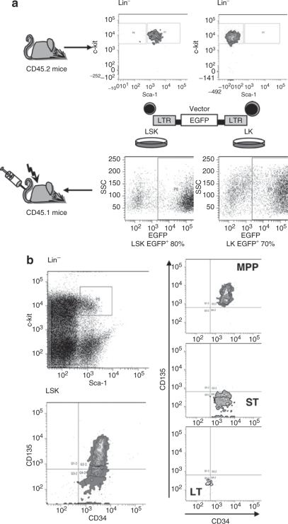
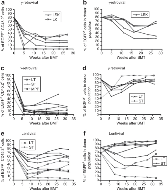
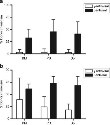
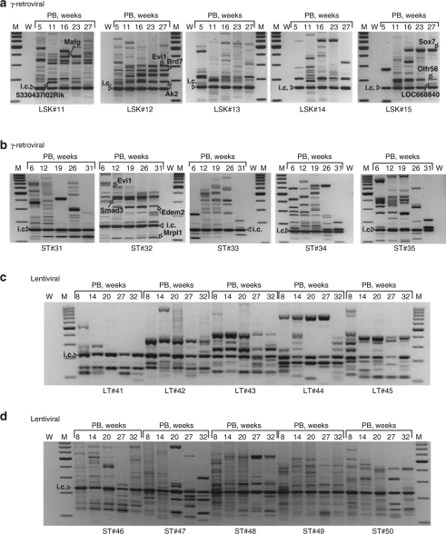
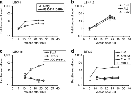
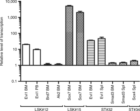
In gene therapeutic approaches targeting hematopoietic cells, insertional mutagenesis may provoke clonal dominance with potential progress to overt leukemia. To investigate the contribution of cell-intrinsic features and determine the frequency of insertional proto-oncogene activation, we sorted hematopoietic subpopulations before transduction with replication-deficient gamma-retroviral vectors and studied the clonal repertoire in transplanted C57BL/6J mice. Progressive clonal dominance only developed in the progeny of populations with intrinsic stem cell potential, where expanding clones with insertional upregulation of proto-oncogenes such as Evi1 were retrieved with a frequency of approximately 10(-4). Longitudinal studies by high-throughput sequencing and locus-specific quantitative PCR showed clones with >50-fold expansion between weeks 5 and 31 after transplantation. In contrast, insertional events in proto-oncogenes did not endow the progeny of multipotent or myeloid-restricted progenitors with the potential for clonal dominance (risk <10(-6)). Transducing sorted hematopoietic stem cells (HSCs) with self-inactivating (SIN) lentiviral vectors in short-term cultures improved chimerism, and although clonal dominance developed, there was no evidence for insertional events in the vicinity of proto-oncogenes as the underlying cause. We conclude that cell-intrinsic properties cooperate with vector-related features to determine the incidence and consequences of insertional mutagenesis. Furthermore, our study offers perspectives for refinement of animal experiments in the assessment of vector-related genotoxicity.


Figures

<b>Figure 1</b>
Figure 1
Scheme of experiment and cell sorting conditions. (a) Sorted LSK and LK cells from bone marrow of untreated CD45.2 mice were prestimulated in serum-free conditions and transduced with γ-retroviral vector expressing EGFP fluorescent protein. Untransduced controls were included for both LSK and LK groups. On day 4 cells were transplanted into lethally irradiated congenic CD45.1 mice along with fresh CD45.1 competitor cells. Mice were observed for 7 months with regular analysis every 5 weeks of transgene expression and clonal dominance status. (b) The sorting procedure for LT and ST fractions of HSC and MPP followed established conditions.23 EGFP, enhanced green fluorescent protein; LSK, lineage marker negative, Sca1+, and c-Kit+; LTR, long terminal repeat; MPP, multipotent progenitor; SSC, side scatter.
<b>Figure 2</b>
Figure 2
Contribution of EGFP+ donor cells to peripheral blood leukocytes and gene marking within the donor population. (a,b) LSK and LK, γ-retroviral transduction. (c,d) STHSC, LTHSC, MPP, γ-retroviral transduction. (e,f) STHSC and LTHSC, lentiviral transduction. Values at week 0 for STHSC and LTHSC correspond to FACS analysis of cultured aliquots performed 7–18 days after transduction. BM, bone marrow; EGFP, enhanced green fluorescent protein; FACS, fluorescence-activated cell sorter; HSC, hematopoietic stem cell; LSK, lineage marker negative, Sca1+, and c-Kit+; LTHSC, long-term repopulating HSC; MPP, multipotent progenitor; PB, peripheral blood; Spl, spleen; STHSC, short-term repopulating HSC.
<b>Figure 3</b>
Figure 3
Donor chimerism (CD45.2+ cells) analysis in recipients transplanted with progeny of γ-retrovirally or lentivirally transduced sorted populations of HSCs. (a) Donor chimerism final analysis in BM, PB, and Spl of recipients transplanted with progeny of γ-retrovirally or lentivirally transduced long-term repopulating fraction of HSCs (LTHSCs). Average donor chimerism calculated for five mice of each experimental group. Error bars indicate 95% confidence intervals. (b) Donor chimerism final analysis in BM, PB, and Spl of recipients transplanted with progeny of γ-retrovirally or lentivirally transduced short-term repopulating fraction of HSCs (STHSCs). Error bars indicate 95% confidence intervals. BM, bone marrow; HSC, hematopoietic stem cell; LTHSC, long-term repopulating HSC; PB, peripheral blood; Spl, spleen; STHSC, short-term repopulating HSC.
<b>Figure 4</b>
Figure 4
Insertion sites analysis at different time points after BMT. (a) γ-Retroviral VIS analysis in PB samples of individual LSK recipients #11–15 obtained by LMPCR. Arrows indicate selected insertion sites. (b) γ-Retroviral VIS analysis in PB samples of individual STHSC recipients ST#31–35. (c) Lentiviral VIS analysis in PB samples of individual LTHSC recipients LT#41–45. (d) Lentiviral VIS analysis in PB samples of STHSC recipients ST#46–50. BMT, bone marrow transplantation; i.c., internal control of LMPCR reaction; LSK, lineage marker negative, Sca1+, and c-Kit+; M, marker; LMPCR, ligation-mediated PCR; LTHSC, long-term repopulating hematopoietic stem cell; PB, peripheral blood; STHSC, short-term repopulating hematopoietic stem cell; VIS, vector insertion site; W, water; weeks, weeks after transplantation.
<b>Figure 5</b>
Figure 5
Locus-specific qPCR analysis of selected clones in γ-retroviral LSK and STHSC progeny. (a–c) Locus-specific qPCR analysis of selected clones in individual recipients of LSK cells. Relative quantification of a target gene amplicon was estimated in comparison to amplicon at early time points, corresponding week 5 for (a) LSK#11, (b) LSK#12, and week 11 for (c) LSK#15. (d) Locus-specific qPCR analysis of selected clones in recipient ST#32. Relative quantification of Evi1 amplicon was estimated in comparison to amplicon at time point 5 weeks after BMT; for Smad3, Edem2, Mrpl1 amplicons in comparison to 11 weeks. Each analysis was performed in triplicate. Error bars indicate standard deviations. BMT, bone marrow transplantation; LSK, lineage marker negative, Sca1+, and c-Kit+; qPCR, quantitative PCR; STHSC, short-term repopulating hematopoietic stem cell.
<b>Figure 6</b>
Figure 6
Transcriptional dysregulation of loci targeted by γ-retroviral insertional mutagenesis. Real-time RT-PCR shows dysregulation of selected targeted alleles in BM, PB, Spl of selected mice (LSK#12, LSK#15, ST#32, and ST#34). The expression level of the control mouse CD45.2 (BM, PB) or CD45.1 (Spl) was set to 1. Mean values of at least three measurements. Error bars indicate 95% confidence intervals. BM, bone marrow; LSK, lineage marker negative, Sca1+, and c-Kit+; PB, peripheral blood; RT, reverse transcription; Spl, spleen.

References

    1. Niwa H. Open conformation chromatin and pluripotency. Genes Dev. 2007;21:2671–2676. - PubMed
    1. Lansdorp PM. Role of telomerase in hematopoietic stem cells. Ann N Y Acad Sci. 2005;1044:220–227. - PubMed
    1. Martinez-Agosto JA, Mikkola HK, Hartenstein V., and , Banerjee U. The hematopoietic stem cell and its niche: a comparative view. Genes Dev. 2007;21:3044–3060. - PubMed
    1. Hacein-Bey-Abina S, Garrigue A, Wang GP, Soulier J, Lim A, Morillon E, et al. Insertional oncogenesis in 4 patients after retrovirus-mediated gene therapy of SCID-X1. J Clin Invest. 2008;118:3132–3142. - PMC - PubMed
    1. Aiuti A, Cattaneo F, Galimberti S, Benninghoff U, Cassani B, Callegaro L, et al. Gene therapy for immunodeficiency due to adenosine deaminase deficiency. N Engl J Med. 2009;360:447–458. - PubMed

Publication types

LinkOut - more resources