

Age-related changes in glutamate release in the CA3 and dentate gyrus of the rat hippocampus
- PMID: 19535175
- PMCID: PMC3407977
- DOI: 10.1016/j.neurobiolaging.2009.05.009
Age-related changes in glutamate release in the CA3 and dentate gyrus of the rat hippocampus
Abstract
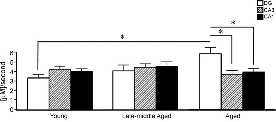
The present studies employed a novel microelectrode array recording technology to study glutamate release and uptake in the dentate gyrus, CA3 and CA1 hippocampal subregions in anesthetized young, late-middle aged and aged male Fischer 344 rats. The mossy fiber terminals in CA3 showed a significantly decreased amount of KCl-evoked glutamate release in aged rats compared to both young and late-middle-aged rats. Significantly more KCl-evoked glutamate release was seen from perforant path terminals in the DG of late-middle-aged rats compared young and aged rats. The DG of aged rats developed an increased glutamate uptake rate compared to the DG of young animals, indicating a possible age-related change in glutamate regulation to deal with increased glutamate release that occurred in late-middle age. No age-related changes in resting levels of glutamate were observed in the DG, CA3 and CA1. Taken together, these data support dynamic changes to glutamate regulation during aging in subregions of the mammalian hippocampus that are critical for learning and memory.
Copyright © 2009 Elsevier Inc. All rights reserved.
Figures


Similar articles
-
The 5-hydroxytryptamine4 receptor enables differentiation of informational content and encoding in the hippocampus.Hippocampus. 2016 Jul;26(7):875-91. doi: 10.1002/hipo.22569. Epub 2016 Feb 17. Hippocampus. 2016. PMID: 26800645 Free PMC article.
-
Regional differences in GABAergic modulation for TEA-induced synaptic plasticity in rat hippocampal CA1, CA3 and dentate gyrus.Neurosci Res. 2007 Oct;59(2):183-90. doi: 10.1016/j.neures.2007.06.1472. Epub 2007 Jun 28. Neurosci Res. 2007. PMID: 17669533
-
Modulation of intracellular calcium changes and glutamate release by neuropeptide Y1 and Y2 receptors in the rat hippocampus: differential effects in CA1, CA3 and dentate gyrus.J Neurochem. 2001 Oct;79(2):286-96. doi: 10.1046/j.1471-4159.2001.00560.x. J Neurochem. 2001. PMID: 11677256
-
Prenatal nicotine and maternal deprivation stress de-regulate the development of CA1, CA3, and dentate gyrus neurons in hippocampus of infant rats.PLoS One. 2013 Jun 13;8(6):e65517. doi: 10.1371/journal.pone.0065517. Print 2013. PLoS One. 2013. PMID: 23785432 Free PMC article.
-
Function of local circuits in the hippocampal dentate gyrus-CA3 system.Neurosci Res. 2019 Mar;140:43-52. doi: 10.1016/j.neures.2018.11.003. Epub 2018 Nov 5. Neurosci Res. 2019. PMID: 30408501 Review.
Cited by
-
The role of glutamatergic pathway between septum and hippocampus in the memory formation.EXCLI J. 2013 Jan 21;12:41-51. eCollection 2013. EXCLI J. 2013. PMID: 27231475 Free PMC article. Review.
-
Reduction of vesicle-associated membrane protein 2 expression leads to a kindling-resistant phenotype in a murine model of epilepsy.Neuroscience. 2012 Jan 27;202:77-86. doi: 10.1016/j.neuroscience.2011.11.055. Epub 2011 Dec 13. Neuroscience. 2012. PMID: 22183055 Free PMC article.
-
Hericium erinaceus Extract Exerts Beneficial Effects on Gut-Neuroinflammaging-Cognitive Axis in Elderly Mice.Biology (Basel). 2023 Dec 28;13(1):18. doi: 10.3390/biology13010018. Biology (Basel). 2023. PMID: 38248449 Free PMC article.
-
Differential levels of glutamate dehydrogenase 1 (GLUD1) in Balb/c and C57BL/6 mice and the effects of overexpression of the Glud1 gene on glutamate release in striatum.ASN Neuro. 2011 Apr 21;3(2):e00057. doi: 10.1042/AN20110005. ASN Neuro. 2011. PMID: 21446915 Free PMC article.
-
Ischemic stroke in the elderly: an overview of evidence.Nat Rev Neurol. 2010 May;6(5):256-65. doi: 10.1038/nrneurol.2010.36. Epub 2010 Apr 6. Nat Rev Neurol. 2010. PMID: 20368741 Review.
References
-
- Adams MM, Smith TD, Moga D, Gallagher M, Wang Y, Wolfe BB, Rapp PR, Morrison JH. Hippocampal dependent learning ability correlates with N-methyl-d-aspartate (NMDA) receptor levels in CA3 neurons of young and aged rats. J. Comp. Neurol. 2001 Apr 2;432(2):230–243. - PubMed
-
- Burmeister JJ, Gerhardt GA. Self-referencing ceramic-based multisite microelectrodes for the detection and elimination of interferences from the measurement ofl-glutamate and other analytes. Anal. Chem. 2001 Mar 5;73:1037–1042. - PubMed
-
- Burmeister JJ, Pomerleau F, Palmer M, Day BK, Huettl P, Gerhardt GA. Improved ceramic-based multisite microelectrode for rapid measurements of l-glutamate in the CNS. J. Neurosci. Meth. 2002 Sep 2;119:163–171. - PubMed
-
- Chawla MK, Barnes CA. Hippocampal granule cells in normal aging: insights from electrophysiological and functional imaging experiments. Prog. Brain Res. 2007;163:661–678. (review). - PubMed
Publication types
MeSH terms
Substances
Grants and funding
LinkOut - more resources
Full Text Sources
Medical
Miscellaneous
