. 2009 Aug;297(2):C459-70.
doi: 10.1152/ajpcell.00201.2009. Epub 2009 Jun 17.

Polarized epithelial cells secrete matriptase as a consequence of zymogen activation and HAI-1-mediated inhibition

Affiliations

Polarized epithelial cells secrete matriptase as a consequence of zymogen activation and HAI-1-mediated inhibition

Jehng-Kang Wang et al. Am J Physiol Cell Physiol. 2009 Aug.

Abstract

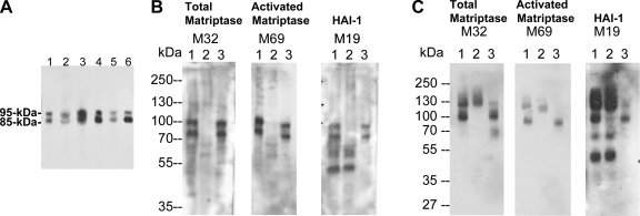
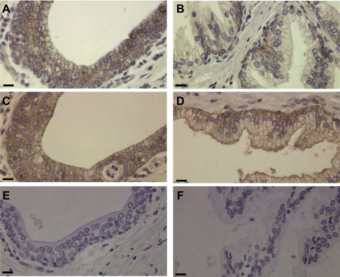
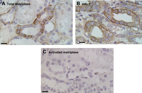
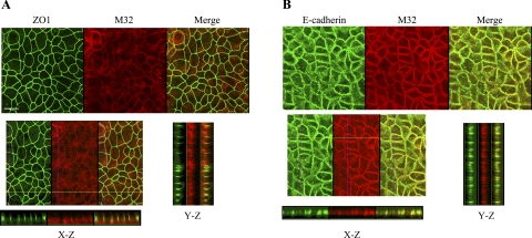
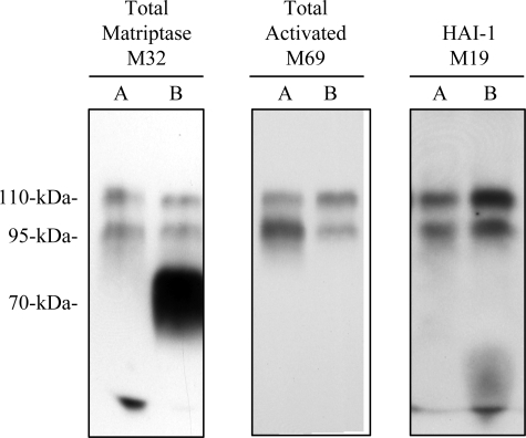
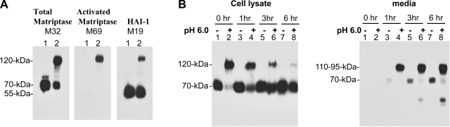
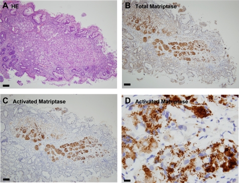
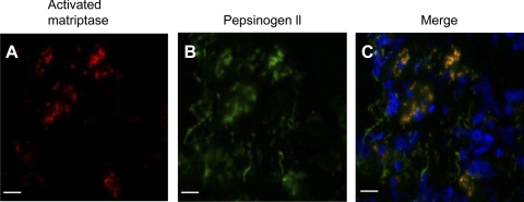
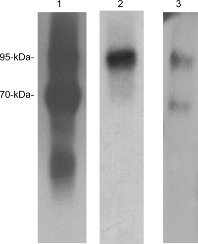
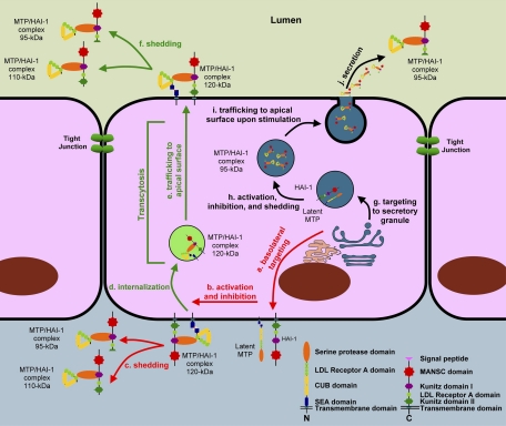
Matriptase, a transmembrane serine protease, is broadly expressed by, and crucial for the integrity of, the epithelium. Matriptase is synthesized as a zymogen and undergoes autoactivation to become an active protease that is immediately inhibited by, and forms complexes with, hepatocyte growth factor activator inhibitor (HAI-1). To investigate where matriptase is activated and how it is secreted in vivo, we determined the expression and activation status of matriptase in seminal fluid and urine and the distribution and subcellular localization of the protease in the prostate and kidney. The in vivo studies revealed that while the latent matriptase is localized at the basolateral surface of the ductal epithelial cells of both organs, only matriptase-HAI-1 complexes and not latent matriptase are detected in the body fluids, suggesting that activation, inhibition, and transcytosis of matriptase would have to occur for the secretion of matriptase. These complicated processes involved in the in vivo secretion were also observed in polarized Caco-2 intestinal epithelial cells. The cells target latent matriptase to the basolateral plasma membrane where activation, inhibition, and secretion of matriptase appear to take place. However, a proportion of matriptase-HAI-1 complexes, but not the latent matriptase, appears to undergo transcytosis to the apical plasma membrane for secretion. When epithelial cells lose their polarity, they secrete both latent and activated matriptase. Although most epithelial cells retain very low levels of matriptase-HAI-1 complex by rapidly secreting the complex, gastric chief cells may activate matriptase and store matriptase-HAI-1 complexes in the pepsinogen-secretory granules, suggesting an intracellular activation and regulated secretion in these cells. Taken together, while zymogen activation and closely coupled HAI-1-mediated inhibition are common features for matriptase regulation, the cellular location of matriptase activation and inhibition, and the secretory route for matriptase-HAI-1 complex may vary along with the functional divergence of different epithelial cells.


Figures

Fig. 1.
Fig. 1.
Secretion of matriptase-hepatocyte growth factor activator inhibitor (HAI-1) complexes, but not latent matriptase in seminal fluid and urine. A: matriptase in human seminal fluid. The presence of matriptase in human seminal fluid from six healthy donors was characterized by immunoblotting using the M32 mAb. Two major bands at around 85 kDa and 95 kDa were observed in all six specimens. B: seminal fluid contains activated matriptase in HAI-1 complexes, but no latent matriptase. The seminal fluid (lanes 1) was subjected to immunoprecipitation with immobilized matriptase mAb 21-9. The unbound fractions were collected (lanes 2), and the matriptase mAb-captured proteins were eluted using acidic buffer, pH 2.4, and then immediately neutralized (lanes 3). All of these samples were analyzed by immunoblotting for total matriptase using the mAb M32 (left, total matriptase, M32), activated matriptase using the mAb M69 (middle, activated matriptase, M69), and HAI-1 using the mAb M19 (right, HAI-1, M19). Both the 85- and the 95-kDa matriptase species were recognized by the activated matriptase mAb and the HAI-1 mAb. Noncomplexed HAI-1 fragment at 50 kDa was also detected by the HAI-1 mAb M19 (right, lane 1) and was not precipitated by matriptase mAb 21–9 beads. C: human urine contains matriptase-HAI-1 complex, but no latent matriptase. The matriptase species present in human urine were assessed by immunoprecipitation followed by immunoblotting through the same procedures as in B. All three mAbs identified the same two proteins bands: one at 95 kDa and a smeared band of >120 kDa (lanes 1). The 95-kDa protein band was depleted by precipitation with matriptase mAb 21-9 beads (lanes 2) and was recognized by the activated matriptase mAb M69 (middle, lane 3) and the HAI-1 mAb M19 (right, lane 3), suggesting that the 95-kDa species is matriptase-HAI-1 complex. The smeared protein bands that were identified in all three immunoblots were not precipitated using the matriptase mAb 21–9 beads (lanes 2), suggesting that the bands do not contain matriptase. The smear of bands is most likely the result of human immunoglobulins in the sample that are identified through cross-reaction with the anti-mouse IgG secondary antibody. A relatively sharp band present at the lower end of the smear (left, lane 1) is, however, immunodepleted by the matriptase mAb 21–9 beads (left, lane 2) and was eluted (left, lane 3). This 110-kDa matriptase band is the matriptase-antithrombin III complex described in our previous study (35). The 50-kDa, free HAI-1 was also detected in human urine by the HAI-1 mAb M19 (right, lane 1) but was not precipitated by the matriptase mAb 21–9 beads, as expected (right, lane 2).
Fig. 2.
Fig. 2.
Expression and distribution of matriptase and HAI-1 in the human prostate. Paraffin-embedded human prostate tissue sections were stained by immunohistochemistry using three mAbs: the M32 for total matriptase (A and B), the M69 for activated matriptase (E and F), and the M19 for HAI-1 (C and D). Positive staining was observed as brown precipitates (diaminobenzidine), and nuclei were counterstained with hematoxylin. The photomicrographs in A, C, and E were taken from the ductal portions of the prostate and in B, D, and F from the acinial portions. Bar = 25 μm.
Fig. 3.
Fig. 3.
Distribution of matriptase and HAI-1 in human kidney. Paraffin-embedded human kidney specimens were stained by immunohistochemistry using mAbs against total matriptase (A), activated matriptase (C), or HAI-1 (B). Positive staining was observed as brown precipitates (diaminobenzidine), and nuclei were counterstained with hematoxylin. Bar = 20 μm.
Fig. 4.
Fig. 4.
Subcellular localization of matriptase in polarized Caco-2 human intestinal epithelial cells. Caco-2 cells were grown and allowed to undergo differentiation on coverslips for 12 days. The polarized cells were fixed, permeabilized, and stained for matriptase with Alexa Fluor 594-conjugated mAb M32. The tight junction marker ZO-1 (A) and the adherens junctions marker E-cadherin (B) were costained with matriptase using FITC-conjugated antibodies to each protein. The staining was observed using a Nikon confocal microscope, and a series of images at different planes in the Z-axis were acquired. Representative images for matriptase, ZO-1, and E-cadherin from both staining studies are shown in the top panels, as indicated. The images in the X-Z- and Y-Z-axes were assembled from serial Z-section images and are shown in the bottom panels, as indicated. The scale bar represents 10 μm.
Fig. 5.
Fig. 5.
Differential secretion of matriptase in polarized Caco-2 human intestinal epithelial cells. Caco-2 cells were grown and allowed to differentiate in transwell chambers for 12 days. The cells were incubated with serum-free culture medium in the top and bottom chambers for 1 day, and the conditioned media from both sides were collected, concentrated, separated by SDS-PAGE, and analyzed by immunoblotting with the matriptase mAb M32, the activated matriptase mAb M69, or the HAI-1 mAb M19, respectively. A, apical surface; B, basolateral surface.
Fig. 6.
Fig. 6.
Matriptase activation is quickly followed by secretion. A: activation and HAI-1-mediated inhibition induced by exposure to a mildly acidic milieu. 184 A1N4 mammary epithelial cells were exposed to basal media or phosphate buffer, pH 6.0, for 20 min. Lysates from both treatments (lanes 1 for nonactivation control; lanes 2 for activation) were subjected to immunoblot analyses for total matriptase using the mAb M32 (left), activated matriptase using the mAb M69 (middle), or HAI-1 using the mAb M19 (right). B: kinetics of matriptase-HAI-1 complex shedding. Matriptase activation was induced by acid exposure, and the cells returned to the basal media for the indicated times. Cell lysates and conditioned media were harvested and analyzed by immunoblotting using the matriptase mAb M32, the activated matriptase mAb M69 (data not shown), and the HAI-1 mAb M19 (data not shown). Control cultures, not exposed to acid, were also examined with the same time course.
Fig. 7.
Fig. 7.
Distribution of matriptase in the human stomach. Paraffin-embedded human stomach sections were stained with hematoxylin and eosin (HE; A) for total matriptase using the mAb S5 (B), and for activated matriptase using the mAb M69 (C and D). The bars are 100 μm in A, B, and C and 10 μm in D.
Fig. 8.
Fig. 8.
Colocalization of activated matriptase with pepsinogen 2 in the secretory granules of gastric chief cells. Paraffin-embedded human stomach sections were stained for activated matriptase (A) and pepsinogen 2 (B) using the mAb M69 and the sheep polyclonal antibody (ab9013), respectively. C: merged image of A and B to show the colocalization between the activated matriptase and pepsinogen 2. Bar = 30 μm.
Fig. 9.
Fig. 9.
Activated matriptase in HAI-1 complexes in human stomach. Normal human stomach tissue was homogenized using RIPA buffer. The lysate was incubated with the matriptase mAb 21-9-Sepharose beads to precipitate total matriptase. The eluted matriptase species were analyzed by immunoblotting for total matriptase using the mAb M32 (lane 1), activated matriptase using the mAb M69 (lane 2), and HAI-1 using the mAb M19 (lane 3).
Fig. 10.
Fig. 10.
The models for matriptase activation, inhibition, and shedding. In most polarized epithelial cells, matriptase is synthesized as a latent zymogen and targeted to basolateral plasma membrane (a). Latent matriptase undergoes autoactivation to generate active matriptase, which is quickly inhibited by HAI-1 to form a 120-kDa complex (b). The 120-kDa matriptase-HAI-1 complex is either shed from the basal plasma membrane (c) or internalized (d) and then traffics to the apical plasma membrane (e) at which the 120-kDa complex is shed into the lumen as 95- and/or 110-kDa complexes by simultaneous proteolytic cleavages at both matriptase and HAI-1 (f). In specific types of epithelial cells, such as the gastric chief cells, matriptase may be synthesized and then targeted to secretory granules (g) within which matriptase undergoes autoactivation, HAI-1-mediated inhibition, and release as a 95-kDa complex with HAI-1, being liberated from the membrane through proteolytic cleavage (h). The 95-kDa matriptase-HAI-1 complex is stored in the secretory granules. Upon stimulation, the secretory granules traffic to apical surface (i), fuse with plasma membrane, and then release the 95-kDa matriptase-HAI-1 complex (j).

Similar articles

Cited by

References

    1. Ahmed S, Jin X, Yagi M, Yasuda C, Sato Y, Higashi S, Lin CY, Dickson RB, Miyazaki K. Identification of membrane-bound serine proteinase matriptase as processing enzyme of insulin-like growth factor binding protein-related protein-1 (IGFBP-rP1/angiomodulin/mac25). FEBS J 273: 615–627, 2006. - PubMed
    1. Benaud C, Dickson RB, Lin CY. Regulation of the activity of matriptase on epithelial cell surfaces by a blood-derived factor. Eur J Biochem 268: 1439–1447, 2001. - PubMed
    1. Benaud C, Oberst M, Hobson JP, Spiegel S, Dickson RB, Lin CY. Sphingosine 1-phosphate, present in serum-derived lipoproteins, activates matriptase. J Biol Chem 277: 10539–10546, 2002. - PubMed
    1. Benaud CM, Oberst M, Dickson RB, Lin CY. Deregulated activation of matriptase in breast cancer cells. Clin Exp Metastasis 19: 639–649, 2002. - PubMed
    1. Bertog M, Letz B, Kong W, Steinhoff M, Higgins MA, Bielfeld-Ackermann A, Fromter E, Bunnett NW, Korbmacher C. Basolateral proteinase-activated receptor (PAR-2) induces chloride secretion in M-1 mouse renal cortical collecting duct cells. J Physiol 521: 3–17, 1999. - PMC - PubMed

Publication types

LinkOut - more resources