

The long-term label retaining population of the renal papilla arises through divergent regional growth of the kidney
- PMID: 19535568
- PMCID: PMC2739716
- DOI: 10.1152/ajprenal.90650.2008
The long-term label retaining population of the renal papilla arises through divergent regional growth of the kidney
Abstract
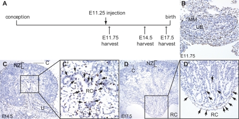
Long-term pulse chase experiments previously identified a sizable population of BrdU-retaining cells within the renal papilla. The origin of these cells has been unclear, and in this work we test the hypothesis that they become quiescent early during the course of kidney development and organ growth. Indeed, we find that BrdU-retaining cells of the papilla can be labeled only by pulsing with BrdU from embryonic (E) day 11.25 to postnatal (P) day 7, the approximate period of kidney development in the mouse. BrdU signal in the cortex and outer medulla is rapidly diluted by cellular proliferation during embryonic development and juvenile growth, whereas cells within the papilla differentiate and exit the cell cycle during organogenesis. Indeed, by E17.5, little or no active proliferation can be seen in the distal papilla, indicating maturation of this structure in a distal-to-proximal manner during organogenesis. We conclude that BrdU-retaining cells of the papilla represent a population of cells that quiesce during embryonic development and localize within a region of the kidney that matures early. We therefore propose that selective papillary retention of BrdU arises through a combination of regionalized slowing of, and exit from, the cell cycle within the papilla during the period of ongoing kidney development, and extensive proliferative growth of the juvenile kidney resulting in dilution of BrdU below the detection level in extra-papillary regions.
Figures


Similar articles
-
Bromodeoxyuridine Methodology for Labeling Renal Stem Cells During Kidney Development of Mice.Stem Cells Dev. 2022 Apr;31(7-8):195-206. doi: 10.1089/scd.2021.0362. Stem Cells Dev. 2022. PMID: 35245977
-
Kinetics of Label Retaining Cells in the Developing Rat Kidneys.PLoS One. 2015 Dec 9;10(12):e0144734. doi: 10.1371/journal.pone.0144734. eCollection 2015. PLoS One. 2015. PMID: 26650841 Free PMC article.
-
The renal papilla is a niche for adult kidney stem cells.J Clin Invest. 2004 Sep;114(6):795-804. doi: 10.1172/JCI20921. J Clin Invest. 2004. PMID: 15372103 Free PMC article.
-
The kidney papilla is a stem cells niche.Stem Cell Rev. 2006;2(3):181-4. doi: 10.1007/s12015-006-0046-3. Stem Cell Rev. 2006. PMID: 17625254 Review.
-
Structural and functional development of outer versus inner cortical proximal tubules.Pediatr Nephrol. 1988 Jan;2(1):108-14. doi: 10.1007/BF00870389. Pediatr Nephrol. 1988. PMID: 3152982 Review.
Cited by
-
Stromal cells in tissue homeostasis: balancing regeneration and fibrosis.Nat Rev Nephrol. 2013 Dec;9(12):747-53. doi: 10.1038/nrneph.2013.152. Epub 2013 Aug 13. Nat Rev Nephrol. 2013. PMID: 23938596 Review.
-
Hyperglycemic Stress Impairs the Stemness Capacity of Kidney Stem Cells in Rats.PLoS One. 2015 Oct 2;10(10):e0139607. doi: 10.1371/journal.pone.0139607. eCollection 2015. PLoS One. 2015. PMID: 26431335 Free PMC article.
-
The role of long-term label-retaining cells in the regeneration of adult mouse kidney after ischemia/reperfusion injury.Stem Cell Res Ther. 2016 Apr 30;7(1):68. doi: 10.1186/s13287-016-0324-1. Stem Cell Res Ther. 2016. PMID: 27137761 Free PMC article.
-
Regenerative medicine for the kidney: stem cell prospects & challenges.Clin Transl Med. 2013 May 21;2(1):11. doi: 10.1186/2001-1326-2-11. Clin Transl Med. 2013. PMID: 23688352 Free PMC article.
-
Repair of injured proximal tubule does not involve specialized progenitors.Proc Natl Acad Sci U S A. 2011 May 31;108(22):9226-31. doi: 10.1073/pnas.1100629108. Epub 2011 May 16. Proc Natl Acad Sci U S A. 2011. PMID: 21576461 Free PMC article.
References
-
- Cha JH, Kim YH, Jung JY, Han KH, Madsen KM, Kim J. Cell proliferation in the loop of Henle in the developing rat kidney. J Am Soc Nephrol 12: 1410–1421, 2001. - PubMed
-
- Cotsarelis G, Cheng SZ, Dong G, Sun TT, Lavker RM. Existence of slow-cycling limbal epithelial basal cells that can be preferentially stimulated to proliferate: implications on epithelial stem cells. Cell 57: 201–209, 1989. - PubMed
-
- Fang TC, Alison MR, Cook HT, Jeffery R, Wright NA, Poulsom R. Proliferation of bone marrow-derived cells contributes to regeneration after folic acid-induced acute tubular injury. J Am Soc Nephrol 16: 1723–1732, 2005. - PubMed
-
- Fujigaki Y, Goto T, Sakakima M, Fukasawa H, Miyaji T, Yamamoto T, Hishida A. Kinetics and characterization of initially regenerating proximal tubules in S3 segment in response to various degrees of acute tubular injury. Nephrol Dial Transplant 21: 41–50, 2006. - PubMed
Publication types
MeSH terms
Substances
Grants and funding
LinkOut - more resources
Full Text Sources