. 2009 Jun 17;29(24):7639-48.
doi: 10.1523/JNEUROSCI.5711-08.2009.

Gephyrin oligomerization controls GlyR mobility and synaptic clustering

Affiliations

Gephyrin oligomerization controls GlyR mobility and synaptic clustering

Martino Calamai et al. J Neurosci. .

Abstract

High local concentrations of glycine receptors (GlyRs) at inhibitory postsynaptic sites are achieved through their binding to the scaffold protein gephyrin. The N- and C-terminal domains of gephyrin are believed to trimerize and dimerize, respectively, thus contributing to the formation of submembranous gephyrin clusters at synapses. GlyRs are associated with gephyrin also at extrasynaptic locations. We have investigated how gephyrin oligomerization influences GlyR dynamics and clustering in COS-7 cells and in cultured spinal cord neurons. To this aim, we have expressed isolated N- and C-terminal domains of gephyrin that interfere with the oligomerization of the full-length protein. We also studied the effect of an endogenous splice variant, ge(2,4,5), with a decreased propensity to trimerize. A reduction of the size and number of gephyrin-GlyR clusters was found in cells expressing the various interfering gephyrin constructs. Using fluorescence recovery after photobleaching, we studied the exchange kinetics of synaptic gephyrin clusters. Real-time single-particle tracking was used to analyze the mobility of GlyRs. We found that all the tested constructs displayed faster rates of recovery than wild-type gephyrin and increased the mobility of extrasynaptic receptors, showing that gephyrin-gephyrin interactions modulate the lateral diffusion of GlyRs. Furthermore, we observed an inverse correlation between GlyR diffusion properties and gephyrin cluster size that depended on the number of binding sites blocked by the different constructs. Since alterations in the oligomerization properties of gephyrin are related to the dynamics of GlyRs, the gephyrin splice variant ge(2,4,5) may be implicated in the modulation of synaptic strength.


Figures
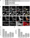
Figure 1.
Figure 1.
Structure and expression pattern of gephyrin constructs in COS-7 cells. A, G, C (or L), and E correspond to the N-terminal, central (or linker), and C-terminal domains of gephyrin, respectively. Ge(2) is the most abundant splice variant of gephyrin. Insertion of cassettes 4 and 5 (light gray boxes) generates the ge(2,4,5) variant. The constructs are N-terminally tagged either with Venus or mrfp. B, COS-7 cells cotransfected with vege(2), mrfpge(2), and the myc-tagged GlyR subunit GlyRα1βgb form gephyrin clusters (both in the green and the red channels) that colocalize with surface GlyRα1βgb immunostained 24 h after transfection. C, Over-expression of the truncated construct mrfpG(2) results in a diffuse distribution and a reduction in the size of gephyrin/GlyRα1βgb coclusters (arrows in the magnified images). Scale bar, 10 μm. D, E, Quantification of the size (D) and relative cellular surface area covered by vege(2)/GlyRα1βgb clusters (E) in COS-7 cells cotransfected with the different mrfp-tagged gephyrin constructs [means ± SEM; N = at least 10 cells for each condition; Student's t test, significant differences from mrfpge(2) with p ≤ 10−3 (***), 10−2 (**), and 5 × 10⁻² (*)].
Figure 2.
Figure 2.
Expression pattern of gephyrin constructs in spinal cord neurons. A, Most of the dendritic vege(2) clusters colocalize with FM4-64-positive presynaptic terminals in live neurons (80.5 ± 3.5%, mean ± SEM, n = 13 cells; 8–10 DIV, 24–48 h after transfection). B, Vege(2) and mrfpge(2) form clusters that colocalize in living neurons. C, The truncated construct mrfpG(2) has a more diffuse distribution and causes a reduction in the size and number of gephyrin clusters (indicated by arrows). Magnified details are shown for each image. Scale bar, 10 μm. D, E, Quantification of the size (D) and the number of vege(2) clusters per length of dendrite (E) in neurons cotransfected with the different mrfp-tagged gephyrin constructs [mean ± SEM; n = 10 cells per condition; Student's t test, significant differences from mrfpge(2) with p ≤ 10−3 (***), 10−2 (**), and 5 × 10⁻² (*)].
Figure 3.
Figure 3.
Dynamics of gephyrin clusters analyzed by FRAP. A, Fluorescence recovery of a gephyrin cluster after photobleaching in a living spinal cord neuron transfected with vege(2) and mrfpge(2). Scale bar, 3 μm. B, Fluorescence recovery of dendritic gephyrin clusters in 8–10 DIV neurons after 24–48 h of expression of vege(2) and mrfpge(2). Venus and mrfp-positive puncta were bleached simultaneously, and the recovery of both constructs was recorded for 30 min. Vege(2) (black circles, black line) and mrfpge(2) (white circles, dotted line) show identical times of recovery. C, The recovery of the truncated construct mrfpG(2) (white circles, dotted line) is almost instantaneous in neurons cotransfected with vege(2) and mrfpG(2), whereas the recovery of vege(2) (black circles, black line) remains unchanged (compare with the black line shown in B and see supplemental Table S1, available at www.jneurosci.org as supplemental material). Fluorescence intensities (mean ± SEM) were normalized to the value before bleaching and to the first value after the bleaching event [with the exception of mrfpG(2); for details, see Materials and Methods]. N > 15 clusters from ≥ 5 cells and >3 independent experiments per condition. D, Endogenous mrfp–gephyrin clusters in spinal cord neurons from a knock-in mouse model (top) colocalize with active presynaptic terminals (FM4-64 staining, bottom image, see arrowheads). Scale bar, 10 μm. E, Fluorescence recovery of mrfp–gephyrin clusters in spinal cord neurons from knock-in animals (mean ± SEM; n = 28 clusters from 7 cells; DIV 10–11). Note that the rate of recovery of endogenous gephyrin is very similar to that of the over-expressed protein (compare with the traces in B).
Figure 4.
Figure 4.
GlyR diffusion in neurons transfected with the various constructs. A, Live imaging of endogenous GlyRs visualized by QDs (red) in spinal cord neurons (DIV 8–10) expressing vege(2) (green). Synapses are identified by the presence of FM4-64 labeling (blue). Receptors are defined as synaptic (s) or extrasynaptic (e) if located over or outside of synapses during the whole duration of the 17.5 s recording session, or as swapping (s/e) if they exchange between the two compartments. Scale bar, 10 μm. B, C, The average MSD of endogenous GlyRs was calculated in synaptic (B) and extrasynaptic locations (C) in neurons transfected with the different Venus-tagged gephyrin constructs. Note the curved MSD plots indicative of the confined motion of synaptic GlyRs (B) and the linear MSD plots of extrasynaptic QDs suggesting Brownian motion (C). D, E, Differences in cumulative fraction of diffusion coefficients were calculated from synaptic (D) and extrasynaptic (E) trajectories, relative to neurons expressing full-length vege(2). Colors in BE correspond to the receptor dynamic in the presence of ge(2) (green), ge(2,4,5) (blue), G(2) (red), G(2,5) (purple), and E (orange). Kolmogorov–Smirnov test: significant differences from vege(2) with p ≤ 10−3 (***) (Table 1).
Figure 5.
Figure 5.
GlyR diffusion in COS-7 cells transfected with the various constructs. A, B, Trajectories of QD-coupled GlyRα1βgb (red) superimposed to binary images of vege(2) clusters (green) in COS-7 cells cotransfected with GlyRα1βgb and vege(2) together with mrfpge(2) (A) or mrfpG(2) (B). Scale bar, 2.5 μm. C, D, Diffusion coefficients of QD-coupled GlyRα1βgb were calculated from trajectories located over (C) and outside of (D) vege(2) clusters in COS-7 cells expressing the different mrfp-tagged constructs. Differences in the cumulative fraction of diffusion coefficients relative to mrfpge(2)-expressing cells show an increased mobility of receptors in the presence of all tested constructs [ge(2,4,5), blue; G(2), red; G(2,5), purple; E, orange; or in the absence of gephyrin, gray]. Kolmogorov–Smirnov test: significant differences from vege(2) with p ≤ 10−3 (***) (see supplemental Table S2, available at www.jneurosci.org as supplemental material).
Figure 6.
Figure 6.
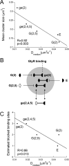
Modification of the gephyrin oligomerization state. A, The reduction of gephyrin cluster size in COS-7 cells in the presence of different interfering constructs is paralleled by an increase in the diffusion of GlyRα1βgb–gephyrin complexes outside of gephyrin clusters. This correlation between receptor diffusion coefficients and cluster size was also observed in neurons (data not shown). B, Schematic representation of the predicted sites of action of the gephyrin constructs used in this study on gephyrin oligomers (synaptic or extrasynaptic) composed of full-length ge(2). Whereas isolated G or E domains interfere with gephyrin oligomerization by saturating binding sites, the full-length ge(2,4,5) variant may replace ge(2) despite its reduced tendency to oligomerize. C, An inverse correlation exists between the diffusion coefficients of GlyRα1βgb outside gephyrin clusters in COS-7 cells and the estimated number of blocked binding sites of the interfering constructs, which appears to define their efficacy.

Similar articles

Cited by

References

    1. Bannai H, Lévi S, Schweizer C, Dahan M, Triller A. Imaging the lateral diffusion of membrane molecules with quantum dots. Nat Protoc. 2006;1:2628–2634. - PubMed
    1. Bedet C, Bruusgaard JC, Vergo S, Groth-Pedersen L, Eimer S, Triller A, Vannier C. Regulation of gephyrin assembly and glycine receptor synaptic stability. J Biol Chem. 2006;281:30046–30056. - PubMed
    1. Bohlhalter S, Mohler H, Fritschy JM. Inhibitory neurotransmission in rat spinal cord: co-localization of glycine- and GABAAreceptors at GABAergic synaptic contacts demonstrated by triple immunofluorescence staining. Brain Res. 1994;642:59–69. - PubMed
    1. Bonneau S, Dahan M, Cohen LD. Single quantum dot tracking based on perceptual grouping using minimal paths in a spatiotemporal volume. IEEE Trans Image Process. 2005;14:1384–1395. - PubMed
    1. Charrier C, Ehrensperger MV, Dahan M, Lévi S, Triller A. Cytoskeleton regulation of glycine receptor number at synapses and diffusion in the plasma membrane. J Neurosci. 2006;26:8502–8511. - PMC - PubMed

Publication types

MeSH terms