Dysfunction of the dentate basket cell circuit in a rat model of temporal lobe epilepsy
- PMID: 19535596
- PMCID: PMC2838908
- DOI: 10.1523/JNEUROSCI.6199-08.2009
Dysfunction of the dentate basket cell circuit in a rat model of temporal lobe epilepsy
Abstract
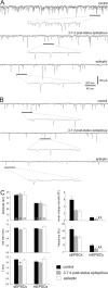
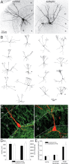
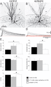
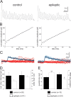
Temporal lobe epilepsy is common and difficult to treat. Reduced inhibition of dentate granule cells may contribute. Basket cells are important inhibitors of granule cells. Excitatory synaptic input to basket cells and unitary IPSCs (uIPSCs) from basket cells to granule cells were evaluated in hippocampal slices from a rat model of temporal lobe epilepsy. Basket cells were identified by electrophysiological and morphological criteria. Excitatory synaptic drive to basket cells, measured by mean charge transfer and frequency of miniature EPSCs, was significantly reduced after pilocarpine-induced status epilepticus and remained low in epileptic rats, despite mossy fiber sprouting. Paired recordings revealed higher failure rates and a trend toward lower amplitude uIPSCs at basket cell-to-granule cell synapses in epileptic rats. Higher failure rates were not attributable to excessive presynaptic inhibition of GABA release by activation of muscarinic acetylcholine or GABA(B) receptors. High-frequency trains of action potentials in basket cells generated uIPSCs in granule cells to evaluate readily releasable pool (RRP) size and resupply rate of recycling vesicles. Recycling rate was similar in control and epileptic rats. However, quantal size at basket cell-to-granule cell synapses was larger and RRP size smaller in epileptic rats. Therefore, in epileptic animals, basket cells receive less excitatory synaptic drive, their pools of readily releasable vesicles are smaller, and transmission failure at basket cell-to-granule cell synapses is increased. These findings suggest dysfunction of the dentate basket cell circuit could contribute to hyperexcitability and seizures.
Figures


Similar articles
-
More Docked Vesicles and Larger Active Zones at Basket Cell-to-Granule Cell Synapses in a Rat Model of Temporal Lobe Epilepsy.J Neurosci. 2016 Mar 16;36(11):3295-308. doi: 10.1523/JNEUROSCI.4049-15.2016. J Neurosci. 2016. PMID: 26985038 Free PMC article.
-
Reduced inhibition of dentate granule cells in a model of temporal lobe epilepsy.J Neurosci. 2003 Mar 15;23(6):2440-52. doi: 10.1523/JNEUROSCI.23-06-02440.2003. J Neurosci. 2003. PMID: 12657704 Free PMC article.
-
Recurrent mossy fiber pathway in rat dentate gyrus: synaptic currents evoked in presence and absence of seizure-induced growth.J Neurophysiol. 1999 Apr;81(4):1645-60. doi: 10.1152/jn.1999.81.4.1645. J Neurophysiol. 1999. PMID: 10200201
-
Mossy fiber-granule cell synapses in the normal and epileptic rat dentate gyrus studied with minimal laser photostimulation.J Neurophysiol. 1999 Oct;82(4):1883-94. doi: 10.1152/jn.1999.82.4.1883. J Neurophysiol. 1999. PMID: 10515977
-
Inhibition of the mammalian target of rapamycin signaling pathway suppresses dentate granule cell axon sprouting in a rodent model of temporal lobe epilepsy.J Neurosci. 2009 Jun 24;29(25):8259-69. doi: 10.1523/JNEUROSCI.4179-08.2009. J Neurosci. 2009. PMID: 19553465 Free PMC article.
Cited by
-
GABA bouton subpopulations in the human dentate gyrus are differentially altered in mesial temporal lobe epilepsy.J Neurophysiol. 2020 Jan 1;123(1):392-406. doi: 10.1152/jn.00523.2018. Epub 2019 Dec 4. J Neurophysiol. 2020. PMID: 31800363 Free PMC article.
-
Initial loss but later excess of GABAergic synapses with dentate granule cells in a rat model of temporal lobe epilepsy.J Comp Neurol. 2010 Mar 1;518(5):647-67. doi: 10.1002/cne.22235. J Comp Neurol. 2010. PMID: 20034063 Free PMC article.
-
Hilar mossy cells of the dentate gyrus: a historical perspective.Front Neural Circuits. 2013 Jan 9;6:106. doi: 10.3389/fncir.2012.00106. eCollection 2012. Front Neural Circuits. 2013. PMID: 23420672 Free PMC article.
-
Persistent decrease in multiple components of the perineuronal net following status epilepticus.Eur J Neurosci. 2012 Dec;36(11):3471-82. doi: 10.1111/j.1460-9568.2012.08268.x. Epub 2012 Aug 31. Eur J Neurosci. 2012. PMID: 22934955 Free PMC article.
-
Basket cell dichotomy in microcircuit function.J Physiol. 2012 Feb 15;590(4):683-94. doi: 10.1113/jphysiol.2011.223669. Epub 2011 Dec 23. J Physiol. 2012. PMID: 22199164 Free PMC article. Review.
References
-
- Andrioli A, Alonso-Nanclares L, Arellano JI, DeFelipe J. Quantitative analysis of parvalbumin-immunoreactive cells in the human epileptic hippocampus. Neuroscience. 2007;149:131–143. - PubMed
-
- Austin JE, Buckmaster PS. Recurrent excitation of granule cells with basal dendrites and low interneuron density and inhibitory postsynaptic current frequency in the dentate gyrus of macaque monkeys. J Comp Neurol. 2004;476:205–218. - PubMed
-
- Babb TL, Kupfer WR, Pretorius JK, Crandall PH, Levesque MF. Synaptic reorganization by mossy fibers in human epileptic fascia dentata. Neuroscience. 1991;42:351–353. - PubMed
Publication types
MeSH terms
Substances
Grants and funding
LinkOut - more resources
Full Text Sources
