. 2009 Oct;81(4):690-9.
doi: 10.1095/biolreprod.109.078261. Epub 2009 Jun 17.

Neonatal bisphenol-a exposure alters rat reproductive development and ovarian morphology without impairing activation of gonadotropin-releasing hormone neurons

Affiliations

Neonatal bisphenol-a exposure alters rat reproductive development and ovarian morphology without impairing activation of gonadotropin-releasing hormone neurons

Heather B Adewale et al. Biol Reprod. 2009 Oct.

Abstract

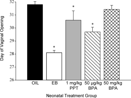
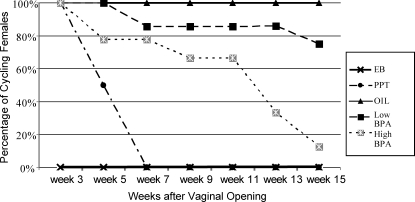
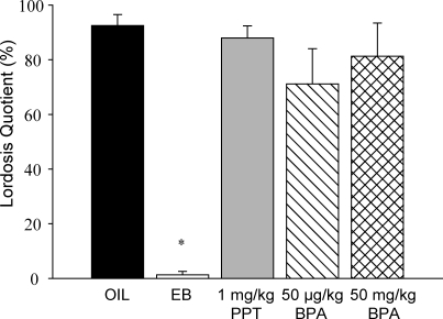
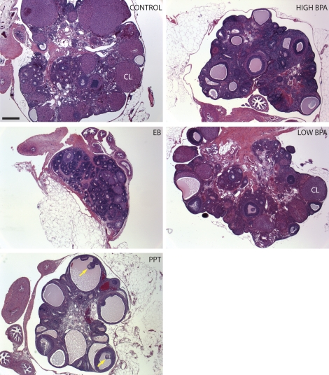
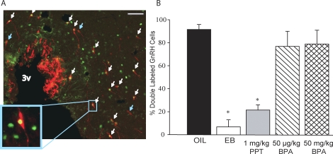
Developmental exposure to endocrine-disrupting compounds is hypothesized to adversely affect female reproductive physiology by interfering with the organization of the hypothalamic-pituitary-gonadal axis. Here, we compared the effects of neonatal exposure to two environmentally relevant doses of the plastics component bisphenol-A (BPA; 50 microg/kg and 50 mg/kg) with the ESR1 (formerly known as ERalpha)-selective agonist 4,4',4''-(4-propyl-[(1)H]pyrazole-1,3,5-triyl)trisphenol (PPT; 1 mg/kg) on the development of the female rat hypothalamus and ovary. An oil vehicle and estradiol benzoate (EB; 25 microg) were used as negative and positive controls. Exposure to EB, PPT, or the low dose of BPA advanced pubertal onset. A total of 67% of females exposed to the high BPA dose were acyclic by 15 wk after vaginal opening compared with 14% of those exposed to the low BPA dose, all of the EB- and PPT-treated females, and none of the control animals. Ovaries from the EB-treated females were undersized and showed no evidence of folliculogenesis, whereas ovaries from the PPT-treated females were characterized by large antral-like follicles, which did not appear to support ovulation. Severity of deficits within the BPA-treated groups increased with dose and included large antral-like follicles and lower numbers of corpora lutea. Sexual receptivity, examined after ovariectomy and hormone replacement, was normal in all groups except those neonatally exposed to EB. FOS induction in hypothalamic gonadotropic (GnRH) neurons after hormone priming was impaired in the EB- and PPT-treated groups but neither of the BPA-treated groups. Our data suggest that BPA disrupts ovarian development but not the ability of GnRH neurons to respond to steroid-positive feedback.


Figures

FIG. 1.
FIG. 1.
The DOV was significantly advanced by neonatal (PND 0 to PND 3) administration of EB and the low (50 μg/kg bw) dose of BPA but not the high (50 mg/kg bw) dose of BPA or the ESR1-selective agonist PPT compared with the control animals (means ± SEM; *P ≤ 0.04).
FIG. 2.
FIG. 2.
Regular estrous cycles commenced in all groups except the group neonatally exposed to EB. The percentage of females within each treatment group displaying a regular estrous cycle diminished over time in all groups except for the control group. By 15 wk after vaginal opening, only 33% of animals exposed to the low (50 μg/kg bw) dose of BPA neonatally still displayed a regular estrous cycle compared with 86% of the animals exposed to the high (50 mg/kg bw) dose of BPA.
FIG. 3.
FIG. 3.
Female sexual receptivity after hormone priming was not significantly affected by neonatal exposure to any of the compounds except for EB (means ± SEM; *P ≤ 0.001).
FIG. 4.
FIG. 4.
Ovarian morphology was distinctly different across treatment groups. Females neonatally exposed to EB had very small ovaries with no clear signs of active oogenesis or ovulation. Ovaries from females neonatally exposed to PPT were characterized by numerous large antral-like follicles, only some of which contained oocytes (yellow arrows). Similar structures were observed in both of the BPA-treated groups but not to the degree seen in females neonatally exposed to PPT. Generally, ovaries from the high-dose (50 mg/kg bw) BPA treatment group more closely resembled those from the PPT treatment group than the EB treatment group. Bar = 500 μm.
FIG. 5.
FIG. 5.
A) Immunolabeling of GnRH (red) and FOS (green) in the OVLT. Double-labeled cells are indicated by the white arrows, and single-labeled GnRH neurons are indicated by the blue arrows. The number of GnRH-immunopositive cells did not significantly differ among groups. Original magnification ×20 (A) and ×63 (inset); bar = 40 μm. B) The percentage of GnRH cells that were colabeled with FOS was significantly lower in animals neonatally exposed to PPT or EB but not significantly affected by neonatal exposure to either the low (50 μg/kg bw) or high (50 mg/kg bw) doses of BPA (means ± SEM; P ≤ 0.001; *P ≤ 0.001). 3v, third ventricle.

References

    1. Dodds EC, Lawson W.Synthetic estrogen agents without the phenanthrene nucleus. Nature 1936; 137: 996
    1. Calafat AM, Kuklenyik Z, Reidy JA, Caudill SP, Ekong J, Needham LL.Urinary concentrations of bisphenol A and 4-nonylphenol in a human reference population. Environ Health Perspect 2005; 113: 391–395.. - PMC - PubMed
    1. Calafat AM, Ye X, Wong LY, Reidy JA, Needham LL.Exposure of the U.S. population to bisphenol A and 4-tertiary-octylphenol: 2003–2004. Environ Health Perspect 2008; 116: 39–44.. - PMC - PubMed
    1. Calafat AM, Weuve J, Ye X, Jia LT, Hu H, Ringer S, Huttner K, Hauser R.Exposure to Bisphenol A and other phenols in neonatal intensive care unit premature infants. Environ Health Perspect 2009; 117: 639–644.. - PMC - PubMed
    1. Tsutsumi O.Assessment of human contamination of estrogenic endocrine-disrupting chemicals and their risk for human reproduction. J Steroid Biochem Mol Biol 2005; 93: 325–330.. - PubMed

Publication types

MeSH terms