. 2009 Jul;68(7):747-56.
doi: 10.1097/NEN.0b013e3181a9d524.

The proapoptotic BH3-only, Bcl-2 family member, Puma is critical for acute ethanol-induced neuronal apoptosis

Affiliations

The proapoptotic BH3-only, Bcl-2 family member, Puma is critical for acute ethanol-induced neuronal apoptosis

Arindam P Ghosh et al. J Neuropathol Exp Neurol. 2009 Jul.

Abstract

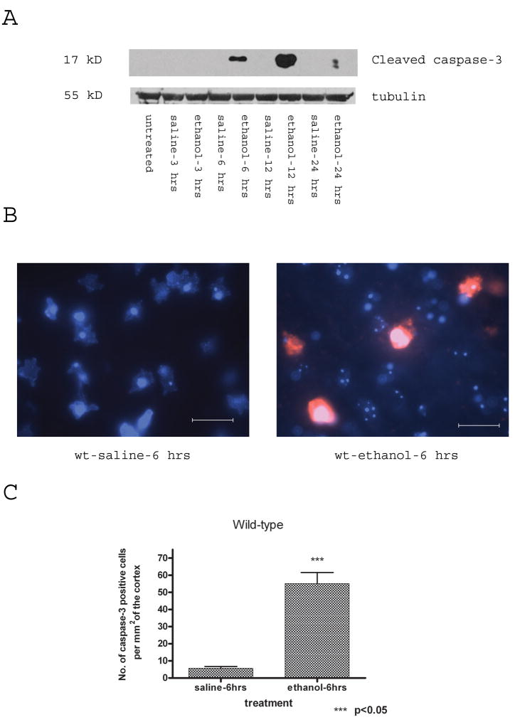
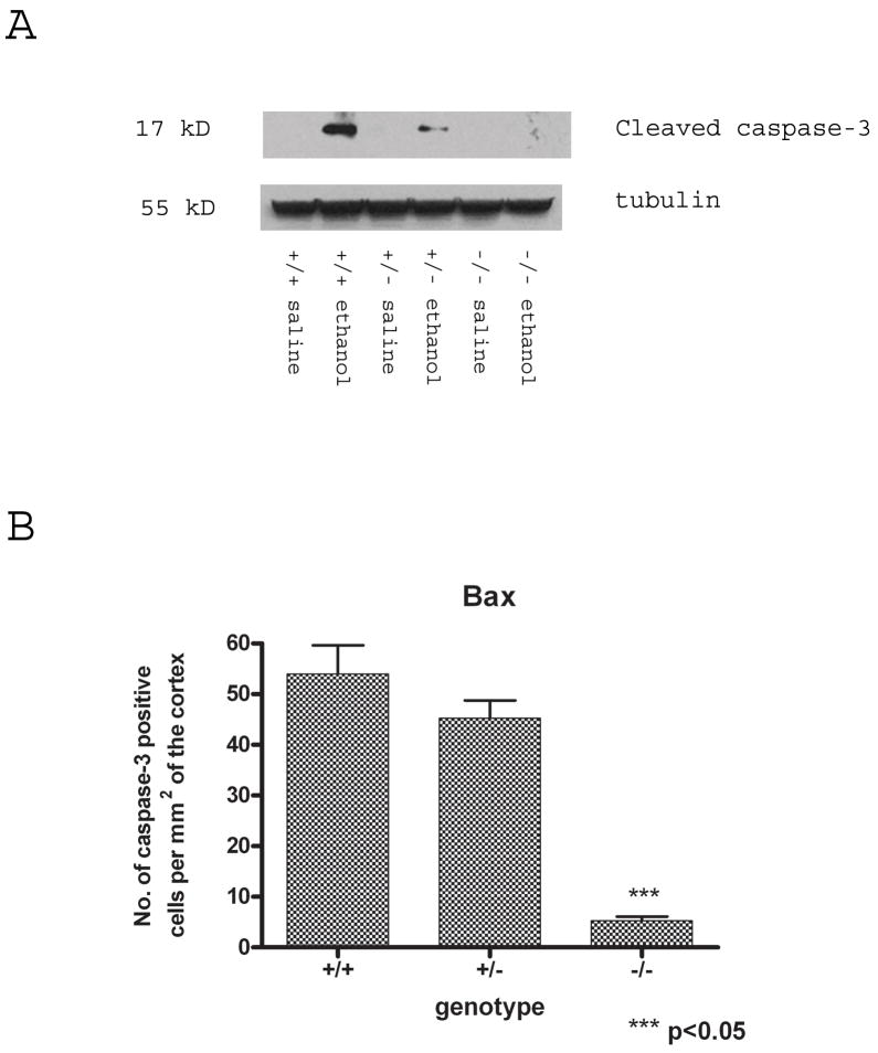
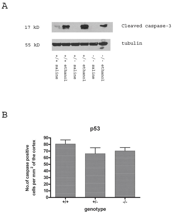
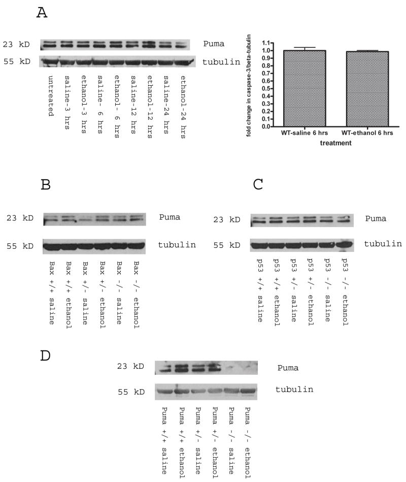
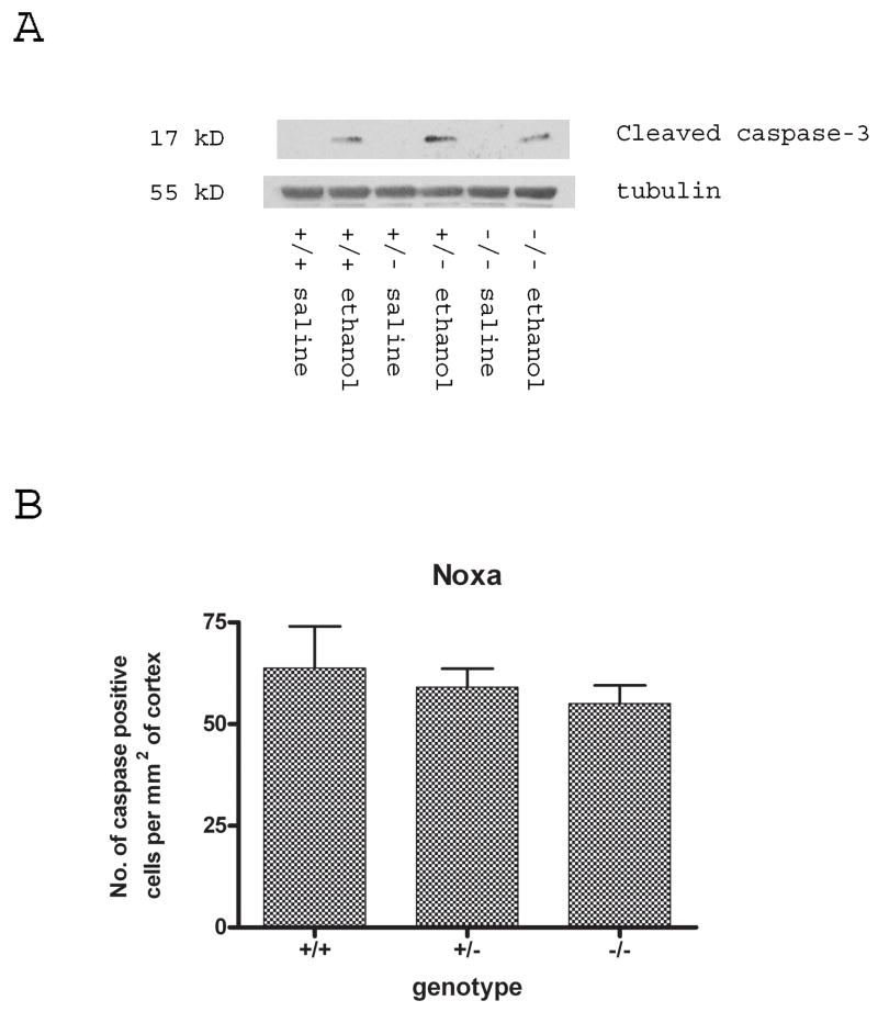
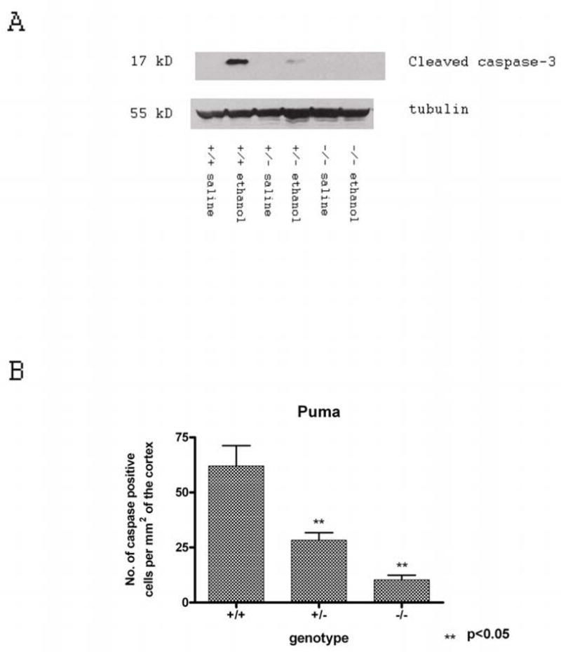
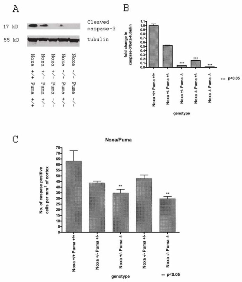
Synaptogenesis in humans occurs in the last trimester of gestation and in the first few years of life, whereas it occurs in the postnatal period in rodents. A single exposure of neonatal rodents to ethanol during this period evokes extensive neuronal apoptosis. Previous studies indicate that ethanol triggers the intrinsic apoptotic pathway in neurons, and that this requires the multi-BH domain, proapoptotic Bcl-2 family member Bax. To define the upstream regulators of this apoptotic pathway, we examined the possible roles of p53 and a subclass of proapoptotic Bcl-2 family members (i.e. the BH3 domain-only proteins) in neonatal wild-type and gene-targeted mice that lack these cell death inducers. Acute ethanol exposure produced greater caspase-3 activation and neuronal apoptosis in wild-type mice than in saline-treated littermate controls. Loss of p53-upregulated mediator of apoptosis (Puma) resulted in marked protection from ethanol-induced caspase-3 activation and apoptosis. Although Puma expression has been reported to be regulated by p53, p53-deficient mice exhibited a similar extent of ethanol-induced caspase-3 activation and neuronal apoptosis as wild-type mice. Mice deficient in other proapoptotic BH3-only proteins, including Noxa, Bim, or Hrk, showed no significant protection from ethanol-induced neuronal apoptosis. Collectively, these studies indicate a p53-independent, Bax- and Puma-dependent mechanism of neuronal apoptosis and identify Puma as a possible molecular target for inhibiting the effects of intrauterine ethanol exposure in humans.


Figures

Figure 1
Figure 1
Postnatal day 7 wild-type pups were injected with ethanol or saline and killed 6 hours later. (A) Western blot analysis of whole brain lysates indicates a time-dependent increase in cleaved (i.e. active) caspase-3 (17 kDa) following ethanol treatment compared to saline-treated time-matched controls. Probing with an antibody specific for β–tubulin was used as a loading control. (B) Immunohistochemical detection of cleaved caspase-3 (Cy3) showed a marked increase in cleaved caspase-3 immunoreactivity in response to ethanol treatment compared to saline-treated littermate controls. Nuclei were counter-stained with bis-benzamide (blue). Scale bar = 50 μm. (C) Ethanol injection induced a significant increase in the number cleaved caspase-3-immunoreactive neurons in the cortex compared to saline-treated neonatal mice. Results are representative of 3 littermate pairs (saline vs. ethanol treated).
Figure 2
Figure 2
Bax deficiency attenuates ethanol-induced caspase-3 activation. (A) Cleaved (i.e. active) caspase-3 was detected by Western blot analysis in whole brain lysates from wild-type and Bax-deficient mice that had been injected with saline or ethanol 6 hours earlier. (B) Bax deficiency significantly protected against ethanol-induced caspase-3 activation in neurons in comparison to wild-type and bax +/− animals. +/+ = wild-type; +/− = heterozygote; −/− = homozygous deficient.
Figure 3
Figure 3
Ethanol-induced caspase-3 cleavage requires Puma expression. Postnatal day 7 pups were injected with ethanol or saline and brains were collected after 6 hours. (A) On Western blot analysis, cleaved caspase-3 was virtually undetectable in brains of saline-treated wild-type mice. Ethanol treatment significantly increased caspase-3 activation in brains of wild-type mice but did not increase cleaved caspase-3 levels in brains from Puma-deficient mice. Ethanol-induced caspase-3 activation was also attenuated in puma+/− mice, suggesting a gene dosage effect. (B) Quantitation of cleaved (i.e., active) caspase-3-immunoreactive cells showed a similar protective effect of loss of Puma in both ethanol-treated puma−/− and puma+/− mice compared to wild-type littermates. Results are representative of 3 littermate pairs (saline vs. ethanol treated). +/+ = wild-type; +/− = heterozygote; −/− = homozygous deficient.
Figure 4
Figure 4
Ethanol-induced caspase-3 activation is dependent upon Puma expression but not p53. (A) Whole brain lysates were prepared from wild-type and p53-deficient mice injected with saline or ethanol and brains were harvested after 6 hours. p53 deficiency did not attenuate ethanol-induced capase-3 activation. (B) Quantification and comparison of the number of cleaved caspase-3-immunoreactive cells between littermates based on their p53 genotypes showed no decrease in numbers of immunoreactive cells in p53−/− animals vs. wild-type and p53+/− littermates. +/+ = wild-type; +/− = heterozygote; −/− = homozygous deficient.
Figure 5
Figure 5
Ethanol treatment does not affect Puma protein levels. (A) Puma protein levels were assessed by Western blotting in the brains of saline- and ethanol-treated wild-type littermates; no significant change was observed. Western blot data are represented graphically (mean ± SEM) as the fold-change in ratio of Puma protein to β-tubulin, ethanol treated relative to saline treated controls. Results are representative of 3 littermate pairs per time point (p < 0.05 versus saline control). (B) Loss of Bax does not affect Puma protein levels in brains of saline- or ethanol-injected mice. Puma protein levels were determined by Western blotting in brains from wild-type, bax+/− or bax−/− mice that had been injected 6 hours earlier with saline or ethanol. (C) Loss of p53 does not affect Puma protein levels in brains of saline- or ethanol-injected mice. Puma protein levels were determined by Western blotting in brains from wild-type, p53+/− or p53−/− mice that had been injected 6 hours earlier with saline or ethanol. (D) Puma protein was not detected by Western blotting in brain extracts from puma −/− mice, demonstrating the specificity of the anti-Puma antibody. An antibody to β-tubulin was used as a loading control in all experiments. +/+ = wild-type; +/− = heterozygote; −/− = homozygous deficient.
Figure 6
Figure 6
Noxa deficiency alone does not protect neurons from the toxic effects of ethanol. (A) The levels of cleaved caspase-3 protein levels determined by Western blotting in brain extracts of wild-type, noxa+/− and noxa−/− mice that had been injected 6 hours earlier with ethanol or saline. Antibody to β-tubulin was used as a loading control. (B) The numbers of cleaved (i.e., active) caspase-3-immunoreactive neurons were determined by immunohistochemistry in brains of wild-type, noxa+/− and noxa−/− mice that had been injected 6 hours earlier with ethanol or saline. +/+ = wild-type; +/− = heterozygote; −/− = homozygous deficient.
Figure 7
Figure 7
Noxa cooperates with Puma in ethanol-induced neuronal cell apoptosis. (A) Cleaved, (i.e., active), caspase-3 levels were determined by Western blotting in brains of wild-type (noxa+/+puma+/+), noxa+/− puma+/−, noxa+/−puma−/−, noxa−/−puma+/− and noxa−/−puma−/− mice that had been injected 6 hours earlier with ethanol or saline. An antibody to β-tubulin was used as a loading control. (B) Quantification of cleaved caspase-3 protein levels by Western blot showed a significant decrease in levels when Noxa deficiency was combined with Puma deficiency; this was significantly lower than the effect of Puma deficiency alone (p < 0.05 by one-way ANOVA). (C) Quantification of cleaved caspase-3-immunoreactive cells showed a significant decrease in the number of caspase-3-immunoreactive cells in noxa+/− puma−/− mice treated with ethanol that was further decreased in the noxa−/− puma−/− mice in response to ethanol (p < 0.05 by one-way ANOVA). +/+ = wild-type; +/− = heterozygote; −/− = homozygous deficient).

Similar articles

Cited by

  • Loss of tumor protein 53 protects against alcohol-induced facial malformations in mice and zebrafish.
    Fish EW, Tucker SK, Peterson RL, Eberhart JK, Parnell SE. Fish EW, et al. Alcohol Clin Exp Res. 2021 Oct;45(10):1965-1979. doi: 10.1111/acer.14688. Epub 2021 Sep 28. Alcohol Clin Exp Res. 2021. PMID: 34581462 Free PMC article.
  • Suppression of the intrinsic apoptosis pathway by synaptic activity.
    Léveillé F, Papadia S, Fricker M, Bell KF, Soriano FX, Martel MA, Puddifoot C, Habel M, Wyllie DJ, Ikonomidou C, Tolkovsky AM, Hardingham GE. Léveillé F, et al. J Neurosci. 2010 Feb 17;30(7):2623-35. doi: 10.1523/JNEUROSCI.5115-09.2010. J Neurosci. 2010. PMID: 20164347 Free PMC article.
  • Gene expression profiling associated with angiotensin II type 2 receptor-induced apoptosis in human prostate cancer cells.
    Pei N, Jie F, Luo J, Wan R, Zhang Y, Chen X, Liang Z, Du H, Li A, Chen B, Zhang Y, Sumners C, Li J, Gu W, Li H. Pei N, et al. PLoS One. 2014 Mar 21;9(3):e92253. doi: 10.1371/journal.pone.0092253. eCollection 2014. PLoS One. 2014. PMID: 24658029 Free PMC article.
  • Caspase-mediated cleavage of actin and tubulin is a common feature and sensitive marker of axonal degeneration in neural development and injury.
    Sokolowski JD, Gamage KK, Heffron DS, Leblanc AC, Deppmann CD, Mandell JW. Sokolowski JD, et al. Acta Neuropathol Commun. 2014 Feb 7;2:16. doi: 10.1186/2051-5960-2-16. Acta Neuropathol Commun. 2014. PMID: 24507707 Free PMC article.
  • Apoptotic cell death in disease-Current understanding of the NCCD 2023.
    Vitale I, Pietrocola F, Guilbaud E, Aaronson SA, Abrams JM, Adam D, Agostini M, Agostinis P, Alnemri ES, Altucci L, Amelio I, Andrews DW, Aqeilan RI, Arama E, Baehrecke EH, Balachandran S, Bano D, Barlev NA, Bartek J, Bazan NG, Becker C, Bernassola F, Bertrand MJM, Bianchi ME, Blagosklonny MV, Blander JM, Blandino G, Blomgren K, Borner C, Bortner CD, Bove P, Boya P, Brenner C, Broz P, Brunner T, Damgaard RB, Calin GA, Campanella M, Candi E, Carbone M, Carmona-Gutierrez D, Cecconi F, Chan FK, Chen GQ, Chen Q, Chen YH, Cheng EH, Chipuk JE, Cidlowski JA, Ciechanover A, Ciliberto G, Conrad M, Cubillos-Ruiz JR, Czabotar PE, D'Angiolella V, Daugaard M, Dawson TM, Dawson VL, De Maria R, De Strooper B, Debatin KM, Deberardinis RJ, Degterev A, Del Sal G, Deshmukh M, Di Virgilio F, Diederich M, Dixon SJ, Dynlacht BD, El-Deiry WS, Elrod JW, Engeland K, Fimia GM, Galassi C, Ganini C, Garcia-Saez AJ, Garg AD, Garrido C, Gavathiotis E, Gerlic M, Ghosh S, Green DR, Greene LA, Gronemeyer H, Häcker G, Hajnóczky G, Hardwick JM, Haupt Y, He S, Heery DM, Hengartner MO, Hetz C, Hildeman DA, Ichijo H, Inoue S, Jäättelä M, Janic A, Joseph B, Jost PJ, Kanneganti TD, Karin M, Kashkar H, Kaufmann T, Kelly … See abstract for full author list ➔ Vitale I, et al. Cell Death Differ. 2023 May;30(5):1097-1154. doi: 10.1038/s41418-023-01153-w. Epub 2023 Apr 26. Cell Death Differ. 2023. PMID: 37100955 Free PMC article. Review.

References

    1. Jones KL, Smith DW. Recognition of the fetal alcohol syndrome in early infancy. Lancet. 1973;2:999–1001. - PubMed
    1. Jones KL, Smith DW, Ulleland CN, et al. Pattern of malformation in offspring of chronic alcoholic mothers. Lancet. 1973;1:1267–71. - PubMed
    1. Clarren SK, Smith DW. The fetal alcohol syndrome. N Engl J Med. 1978;298:1063–67. - PubMed
    1. Swayze VW, Johnson VP, Hanson JW, et al. Magnetic resonance imaging of brain anomalies in fetal alcohol syndrome. Pediatrics. 1997;99:232–40. - PubMed
    1. Streissguth AP, O’Malley K. Neuropsychiatric implications and long-term consequences of fetal alcohol spectrum disorders. Semin Clin Neuropsychiatry. 2000;5:177–90. - PubMed

Publication types

MeSH terms