. 2009 Jul 22;28(14):2028-41.
doi: 10.1038/emboj.2009.162. Epub 2009 Jun 18.

TAK1 is an essential regulator of BMP signalling in cartilage

Affiliations

TAK1 is an essential regulator of BMP signalling in cartilage

Jae-Hyuck Shim et al. EMBO J. .

Abstract

TGFbeta activated kinase 1 (TAK1), a member of the MAPKKK family, controls diverse functions ranging from innate and adaptive immune system activation to vascular development and apoptosis. To analyse the in vivo function of TAK1 in cartilage, we generated mice with a conditional deletion of Tak1 driven by the collagen 2 promoter. Tak1(col2) mice displayed severe chondrodysplasia with runting, impaired formation of secondary centres of ossification, and joint abnormalities including elbow dislocation and tarsal fusion. This phenotype resembled that of bone morphogenetic protein receptor (BMPR)1 and Gdf5-deficient mice. BMPR signalling was markedly impaired in TAK1-deficient chondrocytes as evidenced by reduced expression of known BMP target genes as well as reduced phosphorylation of Smad1/5/8 and p38/Jnk/Erk MAP kinases. TAK1 mediates Smad1 phosphorylation at C-terminal serine residues. These findings provide the first in vivo evidence in a mammalian system that TAK1 is required for BMP signalling and functions as an upstream activating kinase for Smad1/5/8 in addition to its known role in regulating MAP kinase pathways. Our experiments reveal an essential role for TAK1 in the morphogenesis, growth, and maintenance of cartilage.


Conflict of interest statement

The authors declare that they have no conflict of interest.

Figures
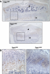
Figure 1
Figure 1
Expression of TAK1 in the proximal tibia. (A) Immunohistochemistry for TAK1 showing expression in a coronal section of the proximal tibias of Tak1fl/fl and Tak1col2 mice. Secondary centres of ossification are indicated with arrows, hypertrophic chondrocytes in the terminal growth plate are indicated with an ‘H'. (B) High power of the indicated areas of (A), showing TAK1 expression in prehypertrophic and hypertrophic chondrocytes.
Figure 2
Figure 2
Abnormal cartilage development of TAK1-deficient mice. (A) Skeletal preparations of 3 week old Tak1fl/fl and Tak1col2 mice (upper panels). X-ray of the skull of 3 week old Tak1fl/fl and Tak1col2 mice, showing doming of the skull and malocclusion in Tak1col2 mice (lower panels). (B) Histological analysis of the proximal tibia of Tak1fl/fl and Tak1col2 mice. Haematoxylin and eosin-stained sections through the proximal tibia of 3 week old Tak1fl/fl and Tak1col2 mice. The proximal tibia of Tak1col2 mice is smaller and shows impaired formation of secondary ossification centres (indicated with an arrow). (C) In situ hybridization for Collagen X〈(ColX) in the proximal humerus of E16.5 and E18.5 Tak1fl/fl and Tak1col2 mice, demonstrating similar extent of chondrocyte hypertrophy. (D) PCNA (top panel) and TUNEL (bottom panel) staining on the chondroepiphyses of p0 Tak1fl/fl and Tak1col2 mice, demonstrating a decrease in PCNA-positive cells and an increase in TUNEL-positive terminal hypertrophic chondrocytes. (E) X-rays and alizarin red/alcian blue stained skeletal preparations of the elbows of 3 week old Tak1fl/fl and Tak1col2 mice, showing dislocation in Tak1col2 mice. (F) Histological analysis of the elbow of Tak1fl/fl and Tak1col2 mice. Haematoxylin and eosin-stained sections through elbows of p20 and E14.5 Tak1fl/fl and Tak1col2 mice. Tak1col2 mice show an overgrowth of the cartilage on the distal humerus and a widening of the proximal radius. The humerus (H), radius (R), and ulna (U) are labelled. (G) High-power view of the ankle of alizarin red/alcian blue stained skeletal preparations of 3 week old Tak1fl/fl and Tak1col2 mice, showing fusion of the tarsal bones and medial dislocation of the phalanges. C, intermediate cuneiform; LC, lateral cuneiform; CU, cuboid; NC, navicular; CA, calcaneus; TA, talus.
Figure 3
Figure 3
Reduced BMP signalling in TAK1-deficient mice. (A) Immunohistochemistry for phosphorylation of BMP-responsive Smad proteins. Coronal sections of the proximal tibia of P0 Tak1fl/fl and Tak1col2 pups were stained with anti-phospho-Smad1/5/8 antibody. Hypertrophic chondrocytes in the terminal growth plate are indicated with an ‘H'. (B) Primary chondrocytes were isolated from cartilage tissues of p0 Tak1fl/fl and Tak1col2 mice, cultured for 3 days, and immunoblotted with the indicated antibodies. (C) In situ hybridization for IHH, Patched, ID1, and Collagen Xα (ColX). Coronal sections of the proximal tibia of 3 week old Tak1fl/fl and Tak1col2 mice were probed for the expression of the indicated mRNAs. The hypertrophic region of the growth plate is shown.
Figure 4
Figure 4
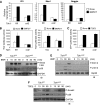
Reduced BMP signalling in TAK1-deficient chondrocytes. (A) Real time-PCR analysis for BMP-responsive gene induction in primary chondrocytes. Tak1fl/fl and Tak1col2 chondrocytes were treated with or without BMP2/7 (100 ng/ml) for 6 h and total RNA was extracted for RT–PCR analysis. (B, C) Immortalized chondrocytes were transfected with either Tlx2-lux (upper panel) or 3TP-luc (lower panel) and Renilla luciferase vectors. Cells were serum starved for 12 h before treatment with BMP2/7 (upper panel) or TGFβ (lower panel), and then analysed for luciferase activity. Results are expressed as relative luciferase activity normalized by Renilla control. (D, E) Immortalized Tak1fl/fl and Tak1col2 chondrocytes were serum starved for 12 h before BMP2/7 (100 ng/ml) stimulation for the indicated times, and then immunoblotted with antibodies specific to phospho-Smad1/5/8 (D), phospho-Erk1/2, or phospho-Jnk1/2 (E). Immunoblotting analysis with antibodies specific to GAPDH (D) or Hsp90 (E) was performed as a control. (F) Immortalized Tak1fl/fl and Tak1col2 chondrocytes were serum starved for 12 h before TGFβ (2 ng/ml) stimulation for the indicated times, and then immunoblotted with antibodies specific to phospho-Smad2 and phospho-p38. Immunoblotting analysis with anti-GAPDH antibody was performed as a control.
Figure 5
Figure 5
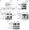
TAK1 is an upstream kinase of Smad proteins in BMP signalling. (A) Direct interaction of TAK1 with BMP-responsive Smad proteins. HEK293 cells were transfected with Flag-Smad1, 5, and 8, and lysed and immunoprecipitated with anti-Flag-conjugated beads. The interactions were tested by Flag pull down assays with Flag-Smad proteins and in vitro translated, 35S-labelled TAK1 (upper panel). Alternatively, GST pull down analysis was performed using either GST or GST-Smad1 and in vitro translated, 35S-labelled TAK1 (lower panel). (B) Endogenous interaction of TAK1 with BMP-responsive Smad proteins. Immortalized chondrocytes were serum starved before BMP2/7 (100 ng/ml) stimulation for the indicated times, and then cells were lysed, immunoprecipitated with either Rabbit IgG or anti-TAK1 antibody and protein A agarose, and immunoblotted with the indicated antibodies. (C) TAK1-induced phosphorylation of Smad proteins. HEK293 cells were transfected with either vector control, constitutively active HA-TAK1 (ΔN), or catalytically inactive HA-TAK1 (CI) and immunoprecipitated with anti-HA antibody and protein G agarose. The immunoprecipitates were mixed with GST, GST-Smad1, Smad2, or Mkk6, and TAK1 kinase activity was analysed by in vitro kinase assay. (D, E) TAK1 activates BMP-responsive Smad1 more efficiently than TGFβ-responsive Smad2. Immortalized wt chondrocytes were serum starved for 12 h before treatment with BMP2/7 (100 ng/ml) or TGFβ (2 ng/ml) for the indicated time and then immunoprecipitated with anti-TAK1 antibody. The immunoprecipitates were mixed with GST-Smad1 (BMP2/7), GST-Smad2 (TGFβ) (D), or GST-Mkk6 (E). TAK1 kinase activity was analysed by in vitro kinase assay.
Figure 6
Figure 6
Identification of Smad1 phosphorylation sites by TAK1. (A) Phosphopeptide analysis of Smad1. HEK293 cells were transfected with Flag-Smad1 together with either vector or HA-TAK1 (ΔN). After immunoprecipitation with anti-Flag conjugated agarose, Smad1 proteins were eluted by Flag peptide, separated by SDS–PAGE, and stained with Coomassie blue (upper panel). The predicted proteolytic digestion pattern of Smad1 (lower panel). (B) Smad1 phosphorylation by TAK1. HEK293 cells were transfected with Flag-Smad1 together with either vector, HA-TAK1 (ΔN), or HA-BMPR1B (Q203D). Cells were lysed and immunoblotted with the indicated antibodies. (C) TAK1-mediated phosphorylation of Smad1 at the C-terminal serines. HEK293 cells were transfected with either vector or HA-TAK1 (ΔN), and the immunoprecipitates were mixed with wt GST-Smad1 (WT) or mutant GST-Smad1 (AAVA). TAK1 kinase activity was analysed by in vitro kinase assay. (D) BMPR1B-mediated phosphorylation of Smad1 at the C-terminal serines. HEK293 cells were transfected with either vector or HA-BMPR1B (Q203D) and the immunoprecipitates were mixed with wt GST-Smad1 (WT) or mutant GST-Smad1 (AAVA). BMPR1B kinase activity was analysed by in vitro kinase assay. (E) Immortalized chondrocytes were serum starved for 12 h before BMP2/7 (100 ng/ml) stimulation for 20 min. The TAK1 immunoprecipitates were washed under stringent conditions to minimize contamination by other endogenous proteins, mixed with GST-Smad1 (WT or AAVA), and TAK1 kinase activity was analysed by in vitro kinase assay.
Figure 7
Figure 7
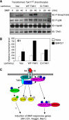
TAK1 kinase activity is important for BMP signalling. (A, B) Immortalized chondrocytes were infected either by vector control, TAK1 (WT), or TAK1 (CI) lentivirues and selected in puromycin-containing medium. (A) Cells were serum starved for 12 h before BMP2/7 (100 ng/ml) stimulation for the indicated time and immunoblotted with antibodies specific to phospho-Smad1/5/8 and phospho-p38. Immunoblotting with anti-Hsp90 and -TAK1 antibodies were performed for protein loading control and TAK1 expression, respectively. (B) Alternatively, cells were unstimulated or stimulated with BMP2/7 for 6 h and total RNA was extracted for RT–PCR analysis. (C) Schematic model of TAK1 function in BMP signalling pathways.

References

    1. Abdollah S, Macias-Silva M, Tsukazaki T, Hayashi H, Attisano L, Wrana JL (1997) TbetaRI phosphorylation of Smad2 on Ser465 and Ser467 is required for Smad2-Smad4 complex formation and signaling. J Biol Chem 272: 27678–27685 - PubMed
    1. Baffi MO, Slattery E, Sohn P, Moses HL, Chytil A, Serra R (2004) Conditional deletion of the TGF-beta type II receptor in Col2a expressing cells results in defects in the axial skeleton without alterations in chondrocyte differentiation or embryonic development of long bones. Dev Biol 276: 124–142 - PubMed
    1. Bandyopadhyay A, Tsuji K, Cox K, Harfe BD, Rosen V, Tabin CJ (2006) Genetic analysis of the roles of BMP2, BMP4, and BMP7 in limb patterning and skeletogenesis. PLoS Genet 2: e216. - PMC - PubMed
    1. Binato R, Alvarez Martinez CE, Pizzatti L, Robert B, Abdelhay E (2006) SMAD 8 binding to mice Msx1 basal promoter is required for transcriptional activation. Biochem J 393(Pt 1): 141–150 - PMC - PubMed
    1. Chang H, Huylebroeck D, Verschueren K, Guo Q, Matzuk MM, Zwijsen A (1999) Smad5 knockout mice die at mid-gestation due to multiple embryonic and extraembryonic defects. Development 126: 1631–1642 - PubMed

Publication types

Substances