. 2009 Jul 22;28(14):2066-76.
doi: 10.1038/emboj.2009.161. Epub 2009 Jun 18.

Cep72 regulates the localization of key centrosomal proteins and proper bipolar spindle formation

Affiliations

Cep72 regulates the localization of key centrosomal proteins and proper bipolar spindle formation

Naoki Oshimori et al. EMBO J. .

Abstract

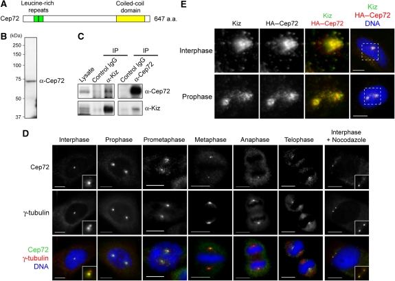
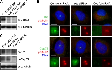
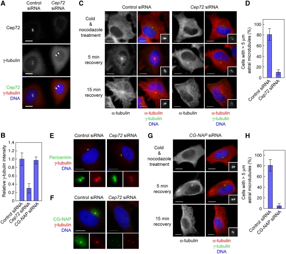
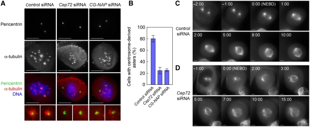
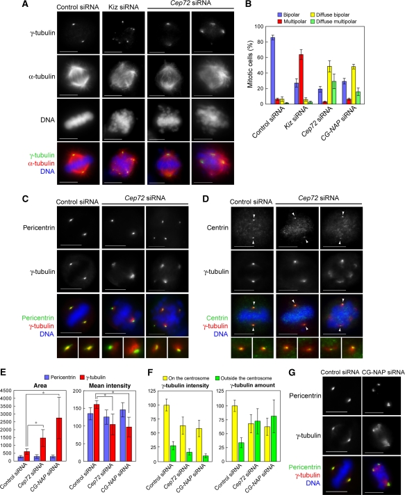
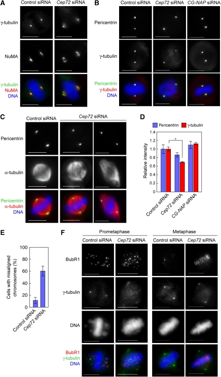
Microtubule-nucleation activity and structural integrity of the centrosome are critical for various cellular functions. The gamma-tubulin ring complexes (gammaTuRCs) localizing to the pericentriolar matrix (PCM) of the centrosome are major sites of microtubule nucleation. The PCM is thought to be created by two cognate large coiled-coil proteins, pericentrin/kendrin and CG-NAP/AKAP450, and its stabilization by Kizuna is essential for bipolar spindle formation. However, the mechanisms by which these proteins are recruited and organized into a proper structure with microtubule-organizing activity are poorly understood. Here we identify a centrosomal protein Cep72 as a Kizuna-interacting protein. Interestingly, Cep72 is essential for the localization of CG-NAP and Kizuna. Cep72 is also involved in gammaTuRC recruitment to the centrosome and CG-NAP confers the microtubule-nucleation activity on the gammaTuRCs. During mitosis, Cep72-mediated microtubule organization is important for converging spindle microtubules to the centrosomes, which is needed for chromosome alignment and tension generation between kinetochores. Our findings show that Cep72 is the key protein essential for maintaining microtubule-organizing activity and structural integrity of the centrosome.


Conflict of interest statement

The authors declare that they have no conflict of interest.

Figures

Figure 1
Figure 1
Cep72 associates and co-localizes with Kiz at and around the centrosome. (A) Schematic diagram of the Cep72 protein. Cep72 has two leucine-rich repeats (green, amino acids (a.a.) 55–76 and 77–98) and a potential coiled-coil domain (yellow, a.a. 476–620). (B) Western blotting of HeLa cell lysates for Cep72. (C) HeLa cell lysates were immunoprecipitated with control IgG or with anti-Cep72 or anti-Kiz antibodies. The co-precipitated proteins were analyzed by immunoblotting using the indicated antibodies. (D) Immunostaining of HeLa cells for Cep72, γ-tubulin, and DNA. (E) To examine the co-localization of Cep72 and Kiz, HeLa cells were transfected with an expression vector for HA–Cep72, fixed, and immunostained for HA (Cep72), Kiz (endogenous), and DNA. Magnified images are of the area within the boxes in the right panels. Scale bar is 10 μm.
Figure 2
Figure 2
Cep72 is required for the centrosomal localization of Kiz. (A) At 60 h after transfection with control or two independent Cep72 siRNAs, HeLa cells were lysed and analyzed by immunoblotting with the indicated antibodies. (B) Control, Kiz, or Cep72 siRNA-transfected HeLa cells were fixed and immunostained for γ-tubulin, DNA, and Kiz (upper panels) or Cep72 (lower panels). Bottom panels of each show enlarged images of the centrosomal area. (C) Control, Kiz, or Cep72 siRNA-transfected cells were lysed and analyzed by immunoblotting using the indicated antibodies. Scale bar is 10 μm.
Figure 3
Figure 3
Cep72 is involved in the centrosomal localization of γTuRC and CG-NAP, and microtubule nucleation from centrosomes. (A) Control or Cep72 siRNA-transfected HeLa cells were plated on the same cover slips, fixed, and immunostained for Cep72, γ-tubulin, and DNA. The arrowheads indicate centrosomes in a Cep72-depleted cell. (B) Comparison of the γ-tubulin intensity of the centrosomes in control, Cep72-depleted, and CG-NAP-depleted cells. To evaluate the γ-tubulin intensity, centrosomal areas were encircled by the edge of γ-tubulin staining and the mean intensity in each area was measured and the background levels subtracted. The data indicate the relative intensity in the centrosomal area and represent the mean±s.d. (n=28). (C) Microtubule re-nucleation assays of control or Cep72 siRNA-transfected interphase cells. After recovering from cold and nocodazole treatment for 0, 5, or 15 min, the cells were fixed and stained for γ-tubulin, α-tubulin, and DNA. Insets show enlargements of the centrosomal area stained for γ-tubulin. (D) The proportion of the length (more than 5 μm) of re-growing microtubules from control or Cep72-depleted centrosomes. Data represent mean±s.d. of three experiments (n>140 for each experiment). (E, F) Control or Cep72-depleted cells were immunostained for γ-tubulin and DNA together with pericentrin (E) or CG-NAP (F). The lower panels show magnified images of the centrosomes. (G) Microtubule re-nucleation assays of CG-NAP siRNA-transfected interphase cells. Compare to the control in (C). (H) The proportion of the length (more than 5 μm) of re-growing microtubules from control or CG-NAP-depleted centrosomes. Data represent mean±s.d. of three experiments (n>100 for each experiment). Scale bar is 10 μm.
Figure 4
Figure 4
Cep72 is required for microtubule aster formation at the onset of mitosis. (A) Microtubule re-growth assays of control, Cep72, or CG-NAP siRNA-transfected mitotic cells. After recovering from cold and nocodazole treatment for 5 min, the cells were fixed and stained for pericentrin, α-tubulin, and DNA. (B) The proportion of control, Cep72-, or CG-NAP-depleted cells with the centrosome-derived microtubule asters at 5 min after re-growth. Data represent the mean±s.d. of three experiments (n>70 for each experiment). (C, D) Time-lapse imaging of mitotic U2OS cells expressing Venus α-tubulin transfected by control (C) or Cep72 siRNA (D). The frame showing NEBD is labelled as 0 min (time in min:s).
Figure 5
Figure 5
Cep72 depletion causes spindle-pole fragmentation and diffuse γTuRC distribution around each spindle pole. (A) Control, Kiz, or Cep72 siRNA-transfected cells were fixed and immunostained for α-tubulin, γ-tubulin, and DNA. (B) The proportion of spindle phenotypes in control, Kiz, Cep72, or CG-NAP siRNA-transfected cells are categorized into four types: bipolar with focused (blue) or diffuse (yellow) γ-tubulin signals, and multi-polar with focused (red) or diffuse (green) γ-tubulin signals. Data represent the mean±s.d. of four experiments (n>100 for each experiment). (C, D) Control or Cep72-depleted HeLa cells were fixed 10–12 h after release from double thymidine block and immunostained for γ-tubulin and DNA, together with pericentrin (a marker for the PCM) (C) or centrin (a centriole marker) (D). The lower panels show magnified images of two spindle poles from each upper panel. Arrowheads indicate the centrioles. (E) Quantification of the area and intensity of pericentrin or γ-tubulin labelling at the poles in bipolar spindles in control, Cep72, and CG-NAP siRNA-treated cells. The stained areas were encircled by the edge of pericentrin or γ-tubulin staining and mean intensities of pericentrin and γ-tubulin in each area were measured. Data show the average intensity in the centrosomal area and represent the mean±s.d. (n>32). *P<0.001. (F) Quantification of the intensity of γ-tubulin signals inside and outside of the pericentrin-staining area. Total amount of γ-tubulin were results from multiplying the values of area and intensity. Data show the average intensities and amounts and the mean±s.d. (n=20). (G) Control or CG-NAP-depleted cells were immunostained for pericentrin, γ-tubulin, and DNA. Scale bar is 10 μm.
Figure 6
Figure 6
Cep72-mediated attachment of spindle microtubules and mitotic centrosomes is required for proper chromosome alignment and segregation. (A) Control or Cep72-depleted cells were fixed and stained for NuMA, γ-tubulin, and DNA. (B) Control, Cep72, or CG-NAP siRNA-transfected cells were incubated in cold media containing 1 μM nocodazole for 30 min to depolymerize microtubules. Cells were fixed and immunostained for pericentrin, γ-tubulin, and DNA. (C) Microtubules were depolymerized as in (B). Mitotic cells were transferred to warm media to allow microtubules to re-grow. After recovering for 30 min, the cells were fixed and immunostained for pericentrin, α-tubulin, and DNA. Pericentrin staining indicates the position of the centrosomes. (D) Quantification of the relative intensity of γ-tubulin labelling at the centrosome in microtubule-depolymerized mitotic cells. Centrosomal areas were encircled by the edge of pericentrin staining and the mean intensities of pericentrin and γ-tubulin in each area were measured. Data show the average intensity in the centrosomal area and represent the mean±s.d. (n=20). *P<0.01. (E) The proportion of cells with misaligned chromosomes in control or Cep72-depleted cells with a bipolar spindle. Data represent the mean±s.d. of four experiments (n>20 for each experiment). (F) Control or Cep72 siRNA transfected HeLa cells were immunostained with BubR1, γ-tubulin, and DNA. Scale bar is 10 μm.

References

    1. Andersen JS, Wilkinson CJ, Mayor T, Mortensen P, Nigg EA, Mann M (2003) Proteomic characterization of the human centrosome by protein correlation profiling. Nature 426: 570–574 - PubMed
    1. Bettencourt-Dias M, Glover DM (2007) Centrosome biogenesis and function: centrosomics brings new understanding. Nat Rev Mol Cell Biol 8: 451–463 - PubMed
    1. Blagden SP, Glover DM (2003) Polar expeditions—provisioning the centrosome for mitosis. Nat Cell Biol 5: 505–511 - PubMed
    1. Bornens M (2002) Centrosome composition and microtubule anchoring mechanisms. Curr Opin Cell Biol 14: 25–34 - PubMed
    1. Casenghi M, Meraldi P, Weinhart U, Duncan PI, Korner R, Nigg EA (2003) Polo-like kinase 1 regulates Nlp, a centrosome protein involved in microtubule nucleation. Dev Cell 5: 113–125 - PubMed

Publication types