

CCR7 signalling as an essential regulator of CNS infiltration in T-cell leukaemia
- PMID: 19536265
- PMCID: PMC3750496
- DOI: 10.1038/nature08020
CCR7 signalling as an essential regulator of CNS infiltration in T-cell leukaemia
Abstract
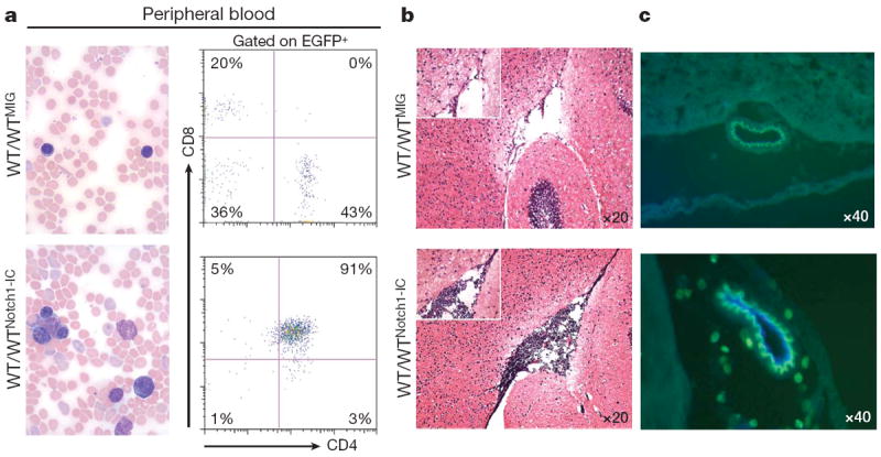
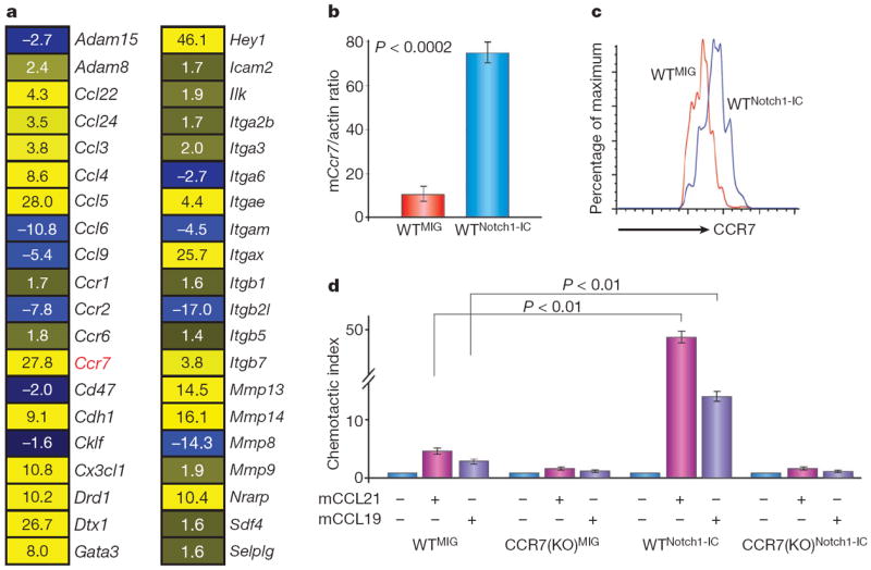
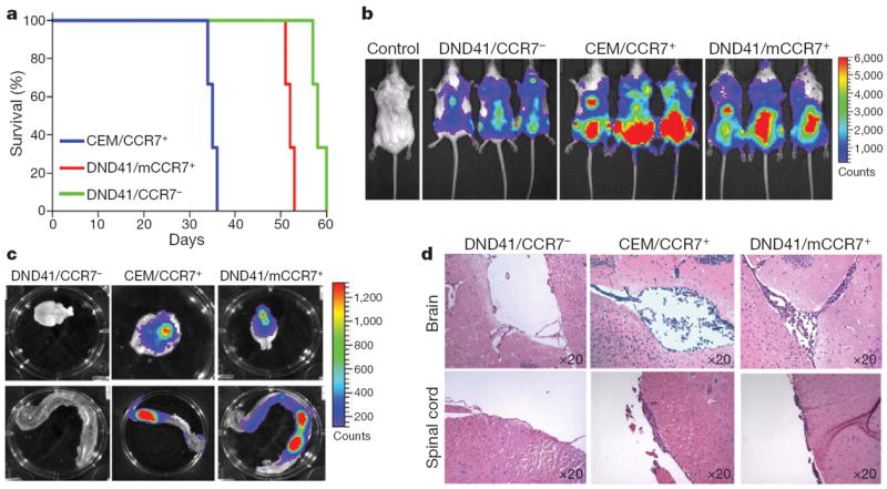
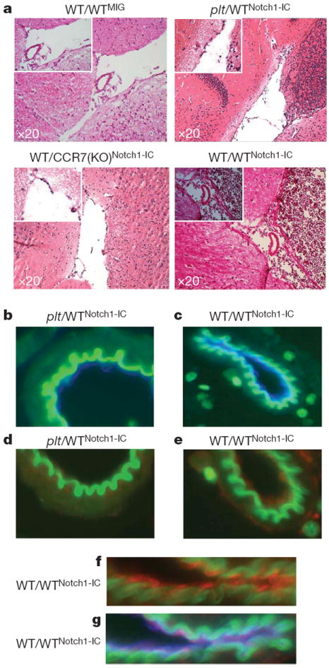
T-cell acute lymphoblastic leukaemia (T-ALL) is a blood malignancy afflicting mainly children and adolescents. T-ALL patients present at diagnosis with increased white cell counts and hepatosplenomegaly, and are at an increased risk of central nervous system (CNS) relapse. For that reason, T-ALL patients usually receive cranial irradiation in addition to intensified intrathecal chemotherapy. The marked increase in survival is thought to be worth the considerable side-effects associated with this therapy. Such complications include secondary tumours, neurocognitive deficits, endocrine disorders and growth impairment. Little is known about the mechanism of leukaemic cell infiltration of the CNS, despite its clinical importance. Here we show, using T-ALL animal modelling and gene-expression profiling, that the chemokine receptor CCR7 (ref. 5) is the essential adhesion signal required for the targeting of leukaemic T-cells into the CNS. Ccr7 gene expression is controlled by the activity of the T-ALL oncogene Notch1 and is expressed in human tumours carrying Notch1-activating mutations. Silencing of either CCR7 or its chemokine ligand CCL19 (ref. 6) in an animal model of T-ALL specifically inhibits CNS infiltration. Furthermore, murine CNS-targeting by human T-ALL cells depends on their ability to express CCR7. These studies identify a single chemokine-receptor interaction as a CNS 'entry' signal, and open the way for future pharmacological targeting. Targeted inhibition of CNS involvement in T-ALL could potentially decrease the intensity of CNS-targeted therapy, thus reducing its associated short- and long-term complications.
Figures
References
-
- Grabher C, von Boehmer H, Look AT. Notch 1 activation in the molecular pathogenesis of T-cell acute lymphoblastic leukaemia. Nature Rev Cancer. 2006;6:347–359. - PubMed
-
- Aifantis I, Raetz E, Buonamici S. Molecular pathogenesis of T-cell leukaemia and lymphoma. Nature Rev Immunol. 2008;8:380–390. - PubMed
-
- Pui CH, Howard SC. Current management and challenges of malignant disease in the CNS in paediatric leukaemia. Lancet Oncol. 2008;9:257–268. - PubMed
-
- Pui CH, Evans WE. Treatment of acute lymphoblastic leukemia. N Engl J Med. 2006;354:166–178. - PubMed
-
- Cyster JG. Chemokines, sphingosine-1-phosphate, and cell migration in secondary lymphoid organs. Annu Rev Immunol. 2005;23:127–159. - PubMed
Publication types
MeSH terms
Substances
Grants and funding
- R01 CA133379/CA/NCI NIH HHS/United States
- R56AI070310/AI/NIAID NIH HHS/United States
- R01CA133379/CA/NCI NIH HHS/United States
- R01 CA149655/CA/NCI NIH HHS/United States
- R01 CA105129/CA/NCI NIH HHS/United States
- R01AI072039/AI/NIAID NIH HHS/United States
- R37 AI062765/AI/NIAID NIH HHS/United States
- R01 AI072039/AI/NIAID NIH HHS/United States
- R21 CA141399/CA/NCI NIH HHS/United States
- R01 AI041428/AI/NIAID NIH HHS/United States
- R01AI41428/AI/NIAID NIH HHS/United States
- R01CA105129/CA/NCI NIH HHS/United States
- 1 P01 CA97403/CA/NCI NIH HHS/United States
- P30 CA016087/CA/NCI NIH HHS/United States
- P01 CA097403/CA/NCI NIH HHS/United States
- R56 AI070310/AI/NIAID NIH HHS/United States
- P30CA016087/CA/NCI NIH HHS/United States
LinkOut - more resources
Full Text Sources
Other Literature Sources
Molecular Biology Databases
