. 2009 Jun 18;4(6):e5968.
doi: 10.1371/journal.pone.0005968.

Prion protein paralog doppel protein interacts with alpha-2-macroglobulin: a plausible mechanism for doppel-mediated neurodegeneration

Affiliations

Prion protein paralog doppel protein interacts with alpha-2-macroglobulin: a plausible mechanism for doppel-mediated neurodegeneration

Stefano Benvegnù et al. PLoS One. .

Erratum in

  • PLoS One. 2009;4(8). doi: 10.1371/annotation/6f581801-c5f0-4fb0-9e55-6ee35653cd12

Abstract

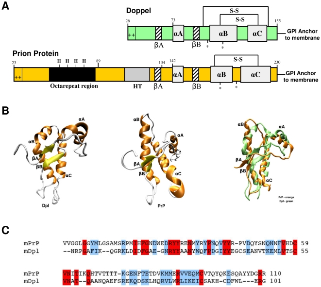
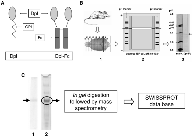
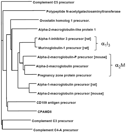
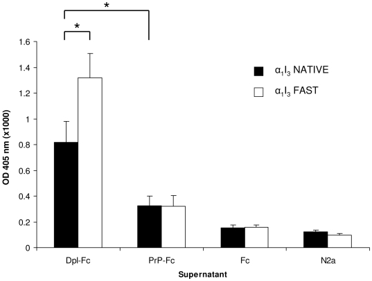
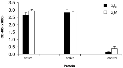
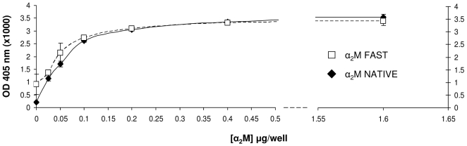
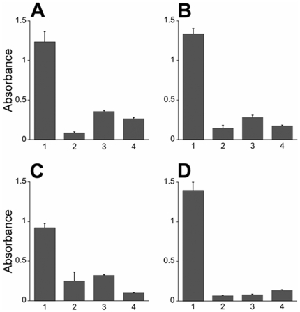
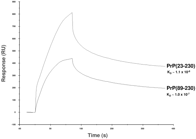
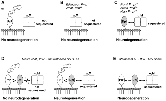
Doppel protein (Dpl) is a paralog of the cellular form of the prion protein (PrP(C)), together sharing common structural and biochemical properties. Unlike PrP(C), which is abundantly expressed throughout the central nervous system (CNS), Dpl protein expression is not detectable in the CNS. Interestingly, its ectopic expression in the brain elicits neurodegeneration in transgenic mice. Here, by combining native isoelectric focusing plus non-denaturing polyacrylamide gel electrophoresis and mass spectrometry analysis, we identified two Dpl binding partners: rat alpha-1-inhibitor-3 (alpha(1)I(3)) and, by sequence homology, alpha-2-macroglobulin (alpha(2)M), two known plasma metalloproteinase inhibitors. Biochemical investigations excluded the direct interaction of PrP(C) with either alpha(1)I(3) or alpha(2)M. Nevertheless, enzyme-linked immunosorbent assays and surface plasmon resonance experiments revealed a high affinity binding occurring between PrP(C) and Dpl. In light of these findings, we suggest a mechanism for Dpl-induced neurodegeneration in mice expressing Dpl ectopically in the brain, linked to a withdrawal of natural inhibitors of metalloproteinase such as alpha(2)M. Interestingly, alpha(2)M has been proven to be a susceptibility factor in Alzheimer's disease, and as our findings imply, it may also play a relevant role in other neurodegenerative disorders, including prion diseases.


Conflict of interest statement

Competing Interests: The authors have declared that no competing interests exist.

Figures

Figure 1
Figure 1. Mature PrP and Dpl protein share common structural architectures.
(A) PrPC and Dpl have common secondary structure elements, composed by three alpha helices (αA, αB and αC) and two beta strands (βA and βB). Both PrPC and Dpl have N-glycosylation sites (*), disulfide bridges (S-S) and a GPI-moiety, which links the proteins to the extracellular side of the cellular membrane. PrPC and Dpl also share a positively charged N-terminus. PrPC contains five octapeptide repeats capable of binding copper through histidine residues (modified from [52]). (B) The topology of Dpl (PDB code: 1I17, left structure) is very similar to that of PrPC (PDB code: 1AG2, central structure). A significant difference is that αB helix of Dpl (in green, right image) is bent and that the two beta strands are oriented differently than those in the PrPC (in orange, right image). (C) Sequence alignment between mouse PrPC (mPrP, residues Val120–Arg229; SwissProt entry: P04925) and mouse Dpl (mDpl, residues Asn55–Gly155; SwissProt entry: Q9QUG3) proteins. In this tract the two proteins share 18% of sequence identity and 44% of sequence similarity. Fully conserved residues are highlighted in red, while semi-conserved are shown in blue.
Figure 2
Figure 2. Identification of Dpl interacting protein in rat cerebellar slices.
(A) Schematic representation of Dpl-Fc construct. Dpl protein is fused with the Fc region of a human immunologlubulin IgG1. The resulting fusion protein is denominated Dpl-Fc. (B) Direct tissue isoelectric focusing of solid tissues technique. This method allows the extraction of more numerous protein vs extracts of tissue homogenates . The technique also prevents the loss of conformational epitopes, which are denatured by standard extraction procedures. Cryostatic rat cerebellar slices (B-1) were put onto the gel. After IEF run, two lateral parts of the gel were blotted onto nitrocellulose stripes, and tested with MoDpl-Fc, for the identification of the binding protein (B-2). Arrows indicate the band (B-2 and B-3). Identification of the band's approximate pI, deduced by comparison with the pH marker, was 5.3. The central part of the gel was used for the protein recovery: a strip of the agarose gel (in white, B-2), which comprised the band (arrows), was excised and placed inside a microtube in PBS. After incubation and centrifugation, the sample was run on native gradient PAGE (C). (C) Gel electrophoresis of recovered protein. To yield amounts of the protein of interest suitable for proteomics, the sample obtained by isoelectric focusing analysis was split into two aliquots and run on PAGE. One aliquot was transferred onto nitrocellulose paper and tested with MoDpl-Fc to identify the protein of interest (C-1), the other one was silver stained (C-2). The arrow indicates the protein that binds to MoDpl-Fc, which was eventually recognized to be rat α1I3 precursor.
Figure 3
Figure 3. Similarity tree of a non-redundant set of the mammalian members of the α2M type of proteinase inhibitors.
Except where noted, the human sequences were taken for alignment purposes. The α2M and rat α1I3 families are respectively highlighted by right braces.
Figure 4
Figure 4. Dpl binds to α1I3.
ELISA plates coated with α1I3 in its native (black bars) or activated, fast (white bars) forms were incubated with the supernatant of respectively MoDpl-Fc transfected N2a cells (Dpl-Fc), MoPrP-Fc transfected N2a cells (PrP-Fc), mock-transfected N2a cells (Fc) and non-transfected N2a cells (N2a). *, p<0.05. Data shown are representative of at least three independent experiments.
Figure 5
Figure 5. ELISA binding measurements of recDpl with α2M and α1I3.
Equimolar amounts of α2M and α1I3 were coated onto ELISA plates both in their native and in their active form, and then incubated with recDpl. Primary antibody was used as control: black bars, α1I3; white bars, α2M. Data shown are representative of at least three independent experiments.
Figure 6
Figure 6. Saturation curve for recDpl with increasing α2M concentrations.
The working range of coated-α2M was from 25 ng to 1.6 µg/well. recDpl was added 1.0 µg/well. A trend of linearity is observed for values up to 100 ng/well, whereas a trend of plateau can be found for values higher than 400 ng/well. No difference is observed in the binding to recDpl comparing the native form with the activated form of α2M. y axis, OD values at 405 nm (x1,000); x axis, µg of proteins per well; white squares, α2M “fast” form; black rhombi, α2M “native” form.
Figure 7
Figure 7. recDpl and recPrP bind to each other.
To determine protein interaction, the primary protein was coated onto a 96-well plate then incubated with the secondary protein. After washing, the presence of the secondary protein was detected by ELISA (Column 1). Control experiments were performed without the primary protein (Column 2), secondary protein (Column 3), or primary antibody (Column 4). (A) Full-length recPrP(23–230) coated onto the plate with recDpl as the secondary protein. Dpl binding was detected using a rabbit polyclonal antibody to Dpl. (B–D) recDpl was coated onto ELISA plates and either recPrP(23–230) (B), recPrP(89–230) (C) or PrP-Fc (D) were incubated and measured for binding. PrP molecules were detected using either a rabbit polyclonal antibody to PrP (B, C) or with an anti-human Fc secondary antibody (D). All ELISA measurements were completed using an AP detection system at 405 nm. y axis, OD values at 405 nm (×1,000). For all panels, graphs represent mean (bar) and standard deviation (error bar) from measurements of at least three independent experiments.
Figure 8
Figure 8. SPR sensorgram comparison of the kinetics of binding between Dpl with either recombinant full-length PrP (PrP(23–230)) or truncated PrP (PrP(89–230)).
Equimolar amounts of PrP(23–230) and PrP(89–230) were injected over Dpl-coupled sensor chip, and kinetics of binding was monitored as response units (RU, y axis) on time (seconds, x axis). Full-length PrP(23–230) proved to possess higher binding capability for Dpl than truncated PrP(89–230). The sensorgrams in the figure were obtained with injection of 800 nM of both PrP(23–230) and PrP(89–230).
Figure 9
Figure 9. Dpl-mediated model of cerebellar neurodegeneration.
The model bears on the postulations as follows: (A) PrP and α2M do not interact each other, therefore α2M is physiologically regulated in the cerebellum; (B) in Prnp 0/0 mice, α2M is still under physiological regulation as in wild type situation; (C) in absence of PrP and in simultaneous presence of Dpl, α2M is sequestered and deregulated, thus leading to cerebellar neurodegeneration. (D) PrP and Dpl are co-expressed, bind and antagonize each other depending on their stoichiometric ratio: on the left, PrP levels are higher than Dpl, PrP sequesters the entire amount of Dpl and thus prevents Dpl interaction with α2M; on the right, Dpl expression is higher than PrP, and residual amounts of Dpl unbound to PrP are still capable of binding α2M. (E) N-terminally truncated PrP binds with less affinity to Dpl, thus permitting it to bind α2M. Mouse models are cited as follows: ZrchI Prnp 0/0 ; Edimburgh Prnp -/- ; Rcm0 Prnp 0/0 ; ZrchII Prnp 0/0 ; Ngsk Prnp 0/0 .

References

    1. Prusiner SB. Prions (Les Prix Nobel Lecture). In: Frängsmyr T, editor. Les Prix Nobel. Stockholm, Sweden: Almqvist & Wiksell International; 1998. pp. 268–323.
    1. Prusiner SB. Shattuck Lecture — Neurodegenerative diseases and prions. N Engl J Med. 2001;344:1516–1526. - PubMed
    1. Moore RC, Lee IY, Silverman GL, Harrison PM, Strome R, et al. Ataxia in prion protein (PrP)-deficient mice is associated with upregulation of the novel PrP-like protein doppel. J Mol Biol. 1999;292:797–817. - PubMed
    1. Silverman GL, Qin K, Moore RC, Yang Y, Mastrangelo P, et al. Doppel is an N-glycosylated, glycosylphosphatidylinositol-anchored protein. Expression in testis and ectopic production in the brains of Prnp(0/0) mice predisposed to Purkinje cell loss. J Biol Chem. 2000;275:26834–26841. - PubMed
    1. Mo H, Moore RC, Cohen FE, Westaway D, Prusiner SB, et al. Two different neurodegenerative diseases caused by proteins with similar structures. Proc Natl Acad Sci U S A. 2001;98:2352–2357. - PMC - PubMed

Publication types