. 2009 Jul 21;42(7):893-903.
doi: 10.1021/ar800245h.

Bioresponsive, cell-penetrating, and multimeric MR contrast agents

Affiliations

Bioresponsive, cell-penetrating, and multimeric MR contrast agents

Jody L Major et al. Acc Chem Res. .

Abstract

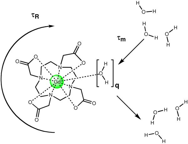
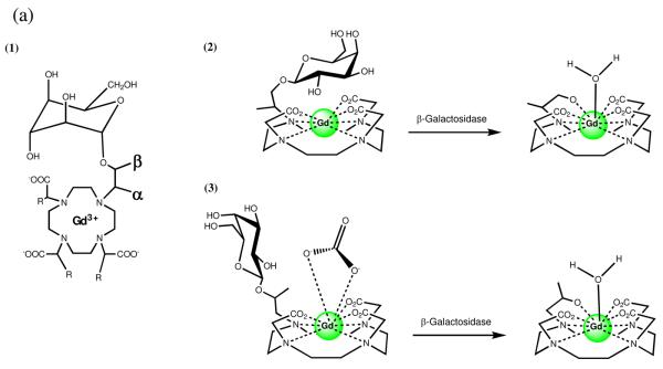
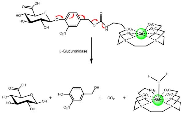
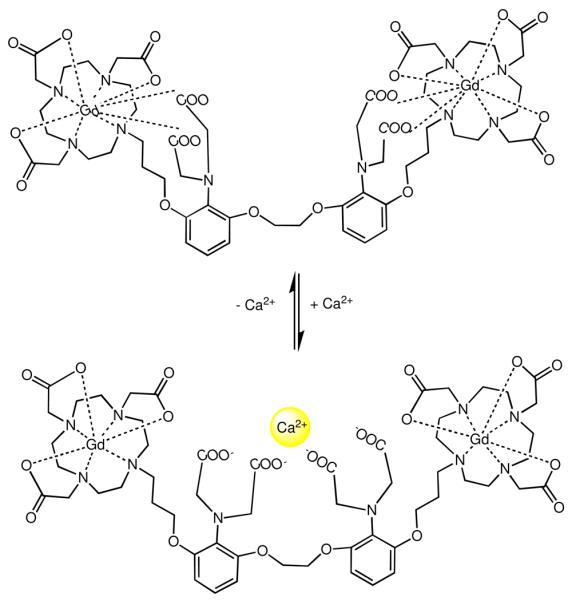
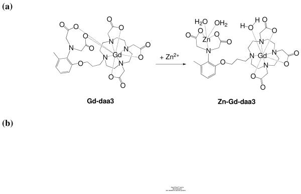
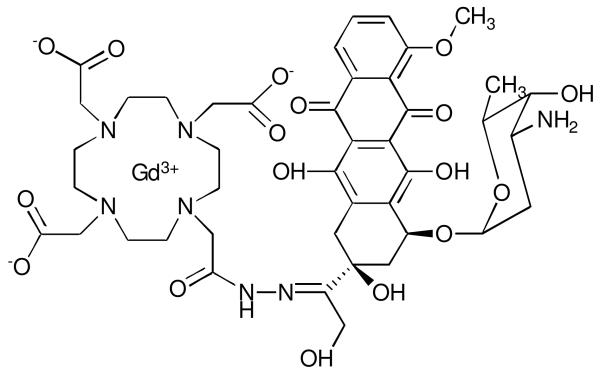
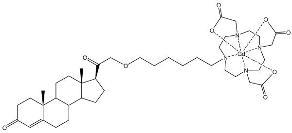
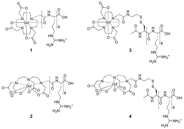
Magnetic resonance imaging (MRI) has become increasingly popular in experimental molecular imaging and clinical radiology because it is non-invasive and capable of producing three-dimensional representations of opaque organisms with high spatial and temporal resolution. Approximately 35% of all clinical MR scans utilize contrast media, however a primary limitation of MR imaging is the sensitivity of contrast agents that require high concentrations (0.1-0.6 mM). A number of strategies have been employed to amplify the observed in vivo signal of MR contrast agents. Approaches include attachment of Gd(III) chelates to polymers, proteins and particles, encapsulation into micelles and caged structures, and targeting to receptors. While each of these approaches has yielded significant increases in the relaxivity of MR contrast agents (and therefore sensitivity), all of these classes of complexes possess intrinsic background signal and function solely as anatomical reporters. In order to reduce the background signal and simultaneously create probes that are modulated by biochemical processes, caged complexes were designed to coordinatively saturate the paramagnetic ion. Coupled with amplification strategies, these agents represent a means to selectively modulate the observed MR signal and function as in vivo biochemical reporters. For example, to create an in vivo MR assay of enzymatic activities and secondary messengers, agents have been designed and synthesized with removable protection groups that largely prevent access of water to a paramagnetic center. By limiting the access of bulk water (q-modulation) the unprocessed agent is designed to be an ineffective contrast agent, and hence serves as a reliable marker for regions of enzyme activity or the presence of secondary messengers. Further, we have focused on designing multimodal contrast agents that are simultaneously detectable by more than one imaging technique. For example, attaching an optical probe to a MR contrast agent provides the means to detect the probe in a whole animal and subsequently validate the results by histological methods. Finally, we describe strategies for signal amplification, and cell delivery vehicles attached to imaging probes for in vivo long-term fate mapping experiments.


Figures

Figure 1
Figure 1
Three important factors that determine the relaxivity of Gd(III)-based MR contrast agents: hydration number (q), the mean residence lifetime of bound water molecules (τm), and the rotational correlation time (τR).
Figure 2
Figure 2
(a) Schematic representations of the two isomers of enzyme-responsive MR agents activated by β-galactosidase: (1) Flat projection with α and β positions labeled (2) α-EGadMe and (3) β-EGadMe. Each isomer provides an increase in relaxivity following cleavage of the sugar by different mechanisms. (b) MRI detection of β-galactosidase mRNA expression in living X. laevis embryos. MR images of two embryos injected with EgadMe at the two-cell stage. (A) Unenhanced MR image. The embryo on the right was injected with β-gal mRNA, resulting in the higher intensity regions. The signal strength is 45–65% greater in the embryo on the right containing β-gal (contrast-to-noise ratio ranges from 3.5 to 6). The cement gland has intrinsically short T1, thus is visible as a bright structure on both embryos. (B) Pseudocolor rendering of same image in (A) with water made transparent. The image correction makes it possible to recognize the eye, and brachial arches in the injected embryo: d, dorsal; v, ventral; r, rostral; e, eye; c, cement gland; s, somite; b, brachial arches. Scale bar = 1 mm.
Figure 2
Figure 2
(a) Schematic representations of the two isomers of enzyme-responsive MR agents activated by β-galactosidase: (1) Flat projection with α and β positions labeled (2) α-EGadMe and (3) β-EGadMe. Each isomer provides an increase in relaxivity following cleavage of the sugar by different mechanisms. (b) MRI detection of β-galactosidase mRNA expression in living X. laevis embryos. MR images of two embryos injected with EgadMe at the two-cell stage. (A) Unenhanced MR image. The embryo on the right was injected with β-gal mRNA, resulting in the higher intensity regions. The signal strength is 45–65% greater in the embryo on the right containing β-gal (contrast-to-noise ratio ranges from 3.5 to 6). The cement gland has intrinsically short T1, thus is visible as a bright structure on both embryos. (B) Pseudocolor rendering of same image in (A) with water made transparent. The image correction makes it possible to recognize the eye, and brachial arches in the injected embryo: d, dorsal; v, ventral; r, rostral; e, eye; c, cement gland; s, somite; b, brachial arches. Scale bar = 1 mm.
Figure 3
Figure 3
A responsive MR contrast agent activated by β-glucuronidase via a self-immolative linker. As with β-EGadMe (Figure 2), endogenous carbonate acts as the ligand to reduce water coordination prior to enzyme activation.
Figure 4
Figure 4
A Ca(II)-activated MR contrast agent Gd-DOPTA. The aminoacetate appended arms internally rearrange upon Ca(II) binding. As a result, a coordination site on the Gd(III) is made available to water (q = 0 to q = 1.5) with a subsequent increase in relaxivity. The agent is selective for Ca(II) only.
Figure 5
Figure 5
(a) Proposed mechanism of a Zn(II)-responsive MR contrast agent. The coordinating acetate arms internally rearrange to bind Zn(II) which opens a coordination site on the Gd(III) center and therefore increases relaxivity. (b) Left: Gd-daa3 Selectivity. T1-weighted MR images of a 1 mM solution of Gd-daa3 in HEPES buffer. (A) HEPES buffer; (B) Gd-daa3; (C) Gd-daa3 with 1 mM ZnCl2; (D) Gd-daa3 with 1 mM MgCl2; (E) Gd-daa3 with 1 mM CaCl2. (b) Right Gd-daa3 Sensitivity. T1-weighted MR images of a 1 mM solution of Gd-daa3 in HEPES buffer with varying zinc concentrations. (A) 0μM Zn; (B) 50 μM Zn; (C) 100 μM Zn; (D) 500 μM Zn; (E) 1 mM Zn.
Figure 6
Figure 6
An example of a prodrug-procontrast complex by conjugating Doxorubicin to a Gd(III) chelate using an acid labile linker. The complex undergoes a change in relaxivity while simultaneously producing an active anticancer drug.
Figure 7
Figure 7
A progesterone Gd(III) chelate conjugate that is designed to image progesterone receptors for early detection of hormone related cancers.
Figure 8
Figure 8
Structures of the arginine-modified (cell-permeable contrast agents) and disulfide bridged (cell-permeable contrast agents) respectively. (1) Gd(III)-DOTA-Arg8, (2) Gd(III)-DTPA-Arg8, (3) Gd(III)-DOTA-SS-Arg8, and (4) Gd(III)-DTPA-SS-Arg8.
Figure 9
Figure 9
T1-weighted MR images of NIH/3T3 cells incubated with complexes 1-4. The ‘No Leach’ rows are the cell populations that were not allowed to leach. The ‘Leach’ rows are the cell populations that were allowed to leach for 4 h with washes of DPBS. The control cells were not incubated with contrast agent but were harvested and packed following the same procedure. (a) Cells incubated with Gd(III)-DOTA-Arg8 or Gd(III)-DTPA-Arg8 (1 and 2). (b) Cells incubated with Gd(III)-DOTA-SS-Arg8 or Gd(III)-DTPA-SS-Arg8 (3 and 4).
Figure 10
Figure 10
(a) Synthetic scheme of multimeric MR contrast agents via click chemistry. (b) The basic structure of labeled PAs containing three sections: a head-group, body, and an alkyl tail. The head-group is composed of an epitope for specific cell interaction and is presented on the outside of the supramolecular fibers.

References

    1. Raymond KN, Pierre VC. Next Generation, High Relaxivity Gadolinium MRI Agents. Bioconjugate Chem. 2005;16:3–8. - PubMed
    1. Lauterbur PC. Image Formation by Incduced Local Interactions: Examples Employing Nuclear Paramagnetic Resonance. Nature. 1973;242:190–191. - PubMed
    1. Merbach A, Toth E. The Chemistry of Contrast Agents in Medical Magnetic Resonance Imaging. John Wiley & Sons, Ltd.; New York: 2001.
    1. Caravan P, Ellison JJ, McMurry TJ, Laufer RB. Gadolinium(III) Chelates as MRI Contrast Agents: Structure, Dynamics, and Applications. Chem. Rev. 1999;99:2293–2352. - PubMed
    1. Solomon I. Relaxation processes in a system of two spins. Physics Reviews. 1955;99:559–565.

Publication types