. 2010 Jun;14(6B):1760-76.
doi: 10.1111/j.1582-4934.2009.00823.x. Epub 2009 Jun 16.

The contact allergen nickel sensitizes primary human endothelial cells and keratinocytes to TRAIL-mediated apoptosis

Affiliations

The contact allergen nickel sensitizes primary human endothelial cells and keratinocytes to TRAIL-mediated apoptosis

Marc Schmidt et al. J Cell Mol Med. 2010 Jun.

Abstract

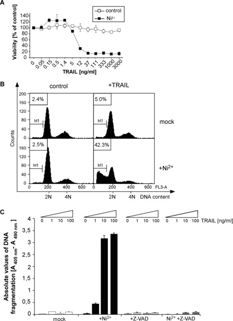
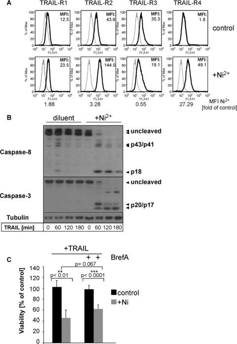
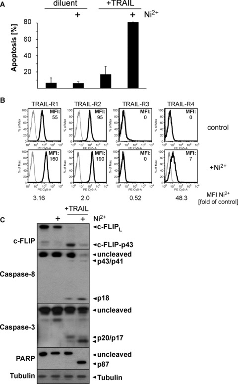
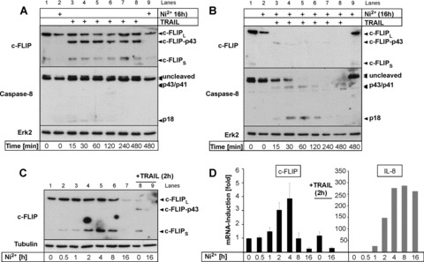
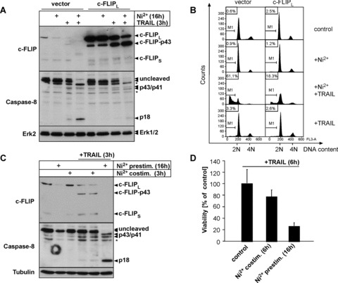
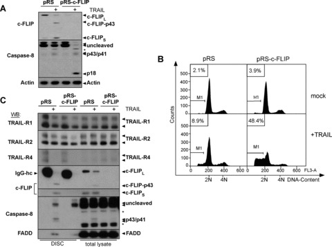
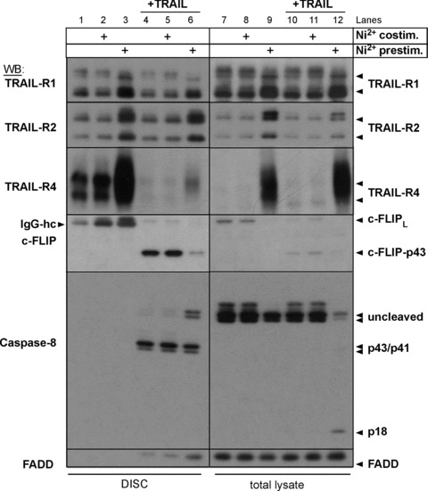
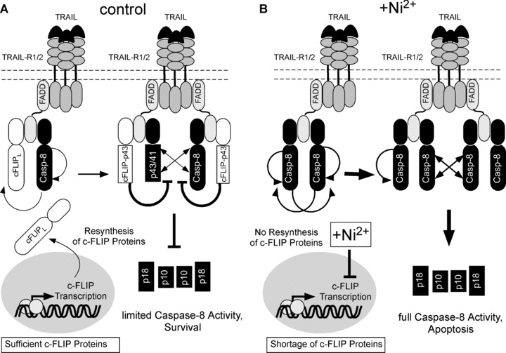
Primary endothelial cells are fully resistant to TNF-related apoptosis-inducing ligand (TRAIL)-mediated apoptosis. Here, we demonstrate that certain environmental conditions, such as exposure to the widespread allergen nickel, can dramatically increase the susceptibility of naturally resistant primary endothelial cells or keratinocytes to TRAIL-induced apoptosis. While nickel treatment increased surface expression of the apoptosis-inducing TRAIL receptors TRAIL-R1 and TRAIL-R2, it also up-regulated the apoptosis-deficient TRAIL-R4, suggesting that modulation of TRAIL receptor expression alone is unlikely to fully account for the dramatic sensitization effect of nickel. Further analysis of candidate mediators revealed that nickel strongly repressed c-FLIP at mRNA and protein levels. Accordingly, increased activation of Caspase-8 and Caspase-3 following nickel treatment was observed. Importantly, depletion of c-FLIP by RNA interference could largely recapitulate the effect of nickel and sensitize endothelial cells to TRAIL-dependent apoptosis in the absence of nickel pre-treatment. Conversely, ectopic expression of c-FLIP(L) largely protected nickel-treated cells from TRAIL-mediated apoptosis. Our data demonstrate that one key mechanism of sensitization of primary human endothelial cells or keratinocytes is transcriptional down-regulation of c-FLIP. We hypothesize that environmental factors, exemplified by the contact allergen nickel, strongly modulate death ligand sensitivity of endothelial cells and keratinocytes thus influencing vascular and epidermal function and integrity under physiological and pathophysiological conditions.


Figures

Fig 1
Fig 1
Ni2+ sensitizes HUVEC to TRAIL-induced apoptosis. (A) Cell viability was assessed by crystal violet staining. HUVEC were left untreated or stimulated with Ni2+ for 16 hrs. Subsequently, cells were cultured for another 6 hrs in Ni2+-free medium containing the indicated TRAIL concentrations. Experimental values were normalized to control values of diluent- or Ni2+-treated controls to facilitate direct comparison. (B) Quantification of apoptosis by flow cytometric analysis of hypodiploid DNA content (M1) of propidium iodide-stained cells. HUVEC were pre-stimulated as in (A) and subsequently exposed to 100 ng/ml TRAIL for 8 hrs. (C) Quantification of apoptosis by measuring internucleosomal DNA fragmentation. HUVEC were treated as in (A) and additionally exposed to 10 μM of the pan-caspase inhibitor zVAD-fmk as indicated. Data in (A) and (C) show mean values ± S.D. from one representative experiment out of 2–3 independent experiments performed in triplicate.
Fig 2
Fig 2
Ni2+ leads to altered TRAIL-receptor expression and rapid Caspase-8 and -3 cleavage upon TRAIL stimulation. (A) Flow cytometric analysis of TRAIL-R1, -R2, -R3 and -R4 cell surface expression of non-stimulated or Ni2+-stimulated (16 hrs) HUVEC using specific antibodies to TRAIL-R1-R4 (filled lines). Grey lines indicate staining with the respective isotype-specific control antibodies. One of four independent experiments is shown with the respective background-subtracted median fluorescence intensity (MFI). Additionally, MFI ratios of Ni2+-treated samples and its respective controls are shown. (B) HUVEC were left untreated or pre-incubated with Ni2+ for 16 hrs and subsequently incubated with TRAIL as indicated. Lysates were analysed for cleavage of Caspase-8 (p43/p41/p18), and Caspase-3 (p20/p17) by Western blot. Membranes were rehybridized with an Ab to tubulin to control for equal protein loading. (C) EC were pre-treated for 16 hrs with Brefeldin A (2 μg/ml) in the presence or absence of Ni2+ (1.5 mM) and subsequently treated with TRAIL (100 ng/ml) for 6 hrs. The viability was subsequently determined by crystal violet assay. Statistical significance of the detected effects was evaluated by Student’s t-test. Statistically significant changes (P < 0.05) are marked by two, highly significant changes (P < 0.005) by three asterisks. Data are derived from a total of five experiments each performed in triplicate wells.
Fig 7
Fig 7
Ni2+ sensitizes human primary keratinocytes for TRAIL-induced apoptosis. Human primary keratinocytes were stimulated with 1.5 mM Ni2+ for 16 hrs or diluent and either directly processed for flow cytometric assessment of TRAIL-R1-4 surface expression (B), or subsequently exposed to 25 ng/ml TRAIL or diluent alone for 6 hrs (A, C). (A) Apoptosis induction as determined by flow cytometric subdiploidy analysis. Average values of two independent experiments using KCs from two different donors ± S.D. are shown. (B) Cell surface expression of TRAIL-R1-4 as determined by flow cytometry. Isotype controls are shown in grey, expression of the respective TRAIL-Rs is depicted in black. Background-corrected MFI values of the stainings are indicated. (C) Western blots showing Ni2+-dependent repression of c-FLIP protein and sensitization of TRAIL-induced apoptosis as evident by induction of Caspase-8, Caspase-3 and PARP cleavage. Tubulin served as a loading control.
Fig 3
Fig 3
c-FLIP is down-regulated by Ni2+ at the mRNA and protein level. (A, B) Untreated HUVEC (A) or HUVEC pre-incubated with Ni2+ for 16 hrs (B) were stimulated with 100 ng/ml TRAIL as indicated and analysed for expression and cleavage of c-FLIP and Caspase-8 by Western blot. Membranes were rehybridized with an Ab to Erk2 to control for even protein loading. Lanes 1, 2 and 9 in (A) and (B) contain equal amounts of identical samples to control for similar exposures of the different membranes. (C, D) HUVEC were treated for the indicated times with Ni2+ and expression of c-FLIP protein (C) or mRNA (D; left panel) was assayed by Western blotting or qRT-PCR, respectively. For comparison, HUVEC were also treated with TRAIL for 2 hrs (C, lanes 8, 9; D, left panel). Data in (D; left panel) represent mean values of two experiments each performed in triplicates ± S.D. In (D; right panel) additional qRT-PCR analysis of IL-8 mRNA for the indicated times is shown.
Fig 4
Fig 4
Transcriptional repression of c-FLIP expression is a functionally relevant mechanism by which Ni2+ sensitizes ECs to TRAIL-induced apoptosis (A, B) Ectopic expression of cFLIPL protects Ni2+-exposed HUVEC against TRAIL-mediated apoptosis. HUVEC were infected with the respective retroviruses and pre-treated with Ni2+ or diluent for 16 hrs. Subsequently, cells were cultured for 3 (A) or 8 hrs (B) in Ni2+-free medium in the presence or absence of TRAIL, respectively. (A) Western blots showing protein expression of c-FLIP and Caspase-8. Molecular weights of full-length proteins and cleavage products are indicated. Asterisks denote unspecific bands consistently obtained with the Caspase-8 antibody. Erk2 served as a loading control. (B) Apoptosis induction as determined by subdiploid DNA analysis. (C, D) Short-term Ni2+-stimulation is insufficient to sensitize ECs to TRAIL-induced apoptosis. HUVEC were either pre-incubated with Ni2+ and subsequently exposed to 100 ng/ml TRAIL or co-stimulated with Ni2+ and TRAIL for 3 (C) or 6 hrs (D). (C) Western blots demonstrate that TRAIL-treated EC maintain c-FLIP protein expression and fail to initiate full Caspase-8 cleavage upon co-treatment of Ni2+ and TRAIL. Tubulin staining served as loading control. (D) Crystal violet viability assays showing strongly decreased capacity of a short-term (6 hrs) Ni2+ co-treatment to sensitize ECs to TRAIL-mediated apoptosis. Data are derived from three independent experiments each performed in triplicate and are presented as percentage of viable cells ± S.D. related to the respective non-Ni2+-stimulated controls that arbitrarily were set to 100%.
Fig 5
Fig 5
Depletion of endogenous c-FLIP by retroviral expression of c-FLIP shRNA sensitizes HUVEC for TRAIL-induced apoptosis. (A–C) HUVEC were infected with the respective constructs and stimulated with 100 ng/ml TRAIL or diluent for 3–8 hrs. (A) Western blot for c-FLIP or Caspase-8 showing TRAIL-dependent cleavage of Caspase-8 upon knock-down of c-FLIP. Molecular weight of full-length proteins and cleavage products are indicated. Cells were lysed 3 hrs after exposure to TRAIL or diluent. The asterisk denotes unspecific bands. Actin served as loading control. (B) Subdiploidy analysis of the differently infected cells harvested 8 hrs after addition of TRAIL or diluent. One representative experiment of three is shown. (C) DISC analysis of TRAIL receptor signalling complexes from the differently infected cells (for details, see Materials and Methods). Cells were cultured for 30 min. in presence of 1 μg/ml Flag-TRAIL pre-complexed with anti-Flag mAb (+) or diluent alone and equal amounts of anti-Flag immunoprecipitates (DISC) were analysed by Western blot. Protein association with non-stimulated receptors was monitored by supplementing lysates from unstimulated cells with anti-Flag-Ab and Flag-TRAIL prior to immunoprecipitation. IgG-hc indicates mouse IgG heavy chain of anti-Flag Ab detected by the respective Abs that is added to the lysates generated from non-stimulated cells as indicated in detail in Materials and Methods. Western blotting with TRAIL-R1, TRAIL-R2 and TRAIL-R4 Abs controls for excessive precipitation of unstimulated receptors as compared to stimulated (+) receptors. Asterisk denotes unspecific bands. Arrows indicate detection of cleavage fragments of the respective proteins.
Fig 6
Fig 6
Prolonged Ni2+-treatment triggers TRAIL-dependent activation of Caspase-8 by reduced c-FLIP recruitment to the TRAIL DISC. EC were pre-treated with Ni2+ for 16 hrs (lanes 3, 6, 9, 12) or treated with diluent alone. Cells were subsequently cultured in the presence of 2.5 μg/ml Flag-TRAIL pre-complexed with anti-Flag mAb (+) or diluent alone in the absence or presence of Ni2+ for 30 min. Subsequently, equal amounts (1 mg of total cellular proteins) were subjected to anti-Flag immunoprecipitation (DISC, lanes 1–6). Total cellular lysates of all conditions were analysed in parallel (total lysate, lanes 7–12). Protein association with non-stimulated receptors was monitored by supplementing equal amounts of lysates from unstimulated cells with Flag-TRAIL that was pre-complexed with anti-Flag mAb prior to immunoprecipitation. IgG-hc indicates mouse IgG heavy chain of anti-Flag mAbs detected by the respective Abs.
Fig 8
Fig 8
Model for Ni2+-dependent regulation of TRAIL resistance in primary human ECs. (A) In the absence of Ni2+, TRAIL stimulation leads to efficient recruitment of FADD, Caspase-8 and c-FLIP to the DISC. c-FLIPL and Caspase-8 are cleaved within the DISC (see arrows), but c-FLIP-p43 prevents further cleavage within and release of Caspase-8 from the DISC. Continuous transcription of c-FLIP proteins maintains blockade of TRAIL death receptors and thus inhibits the DISC turnover over time. (B) In the presence of Ni2+, re-supply of c-FLIP protein is shut off due to repression of c-FLIP transcription. TRAIL stimulation under these conditions leads to an increased caspase-8 recruitment to, cleavage of, and release from the TRAIL DISC thus leading to rapid consumption of remnant c-FLIP protein. Once the cellular pool of c-FLIP is consumed by continuous receptor triggering, Caspase-8 is cleaved in large amounts at the DISC and rapidly released in large quantities, thereby inducing cellular apoptosis. Ni2+-dependent up-regulation of TRAIL-R1 and 2 may further increase the requirement of protective c-FLIP protein at the DISC.

Similar articles

Cited by

References

    1. Krob HA, Fleischer AB, Jr, D’Agostino R, Jr, et al. Prevalence and relevance of contact dermatitis allergens: a meta-analysis of 15 years of published T.R.U.E. test data. J Am Acad Dermatol. 2004;51:349–53. - PubMed
    1. Larese F, Gianpietro A, Venier M, et al. In vitro percutaneous absorption of metal compounds. Toxicol Lett. 2007;170:49–56. - PubMed
    1. Koster R, Vieluf D, Kiehn M, et al. Nickel and molybdenum contact allergies in patients with coronary in-stent restenosis. Lancet. 2000;356:1895–7. - PubMed
    1. Hostynek JJ. Sensitization to nickel: etiology, epidemiology, immune reactions, prevention, and therapy. Rev Environ Health. 2006;21:253–80. - PubMed
    1. Goebeler M, Roth J, Brocker EB, et al. Activation of nuclear factor-kappa B and gene expression in human endothelial cells by the common haptens nickel and cobalt. J Immunol. 1995;155:2459–67. - PubMed

Publication types

MeSH terms