. 2009 Sep 15;47(6):767-78.
doi: 10.1016/j.freeradbiomed.2009.06.017. Epub 2009 Jun 17.

Role of peroxisome proliferator-activated receptor-alpha in fasting-mediated oxidative stress

Affiliations

Role of peroxisome proliferator-activated receptor-alpha in fasting-mediated oxidative stress

Mohamed A Abdelmegeed et al. Free Radic Biol Med. .

Abstract

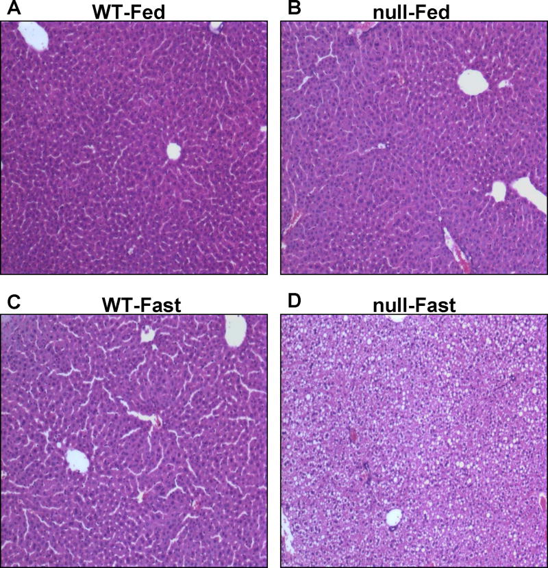
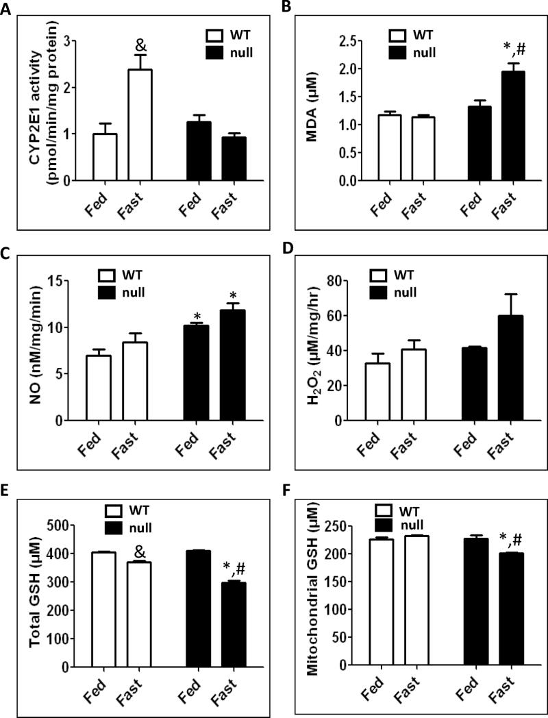
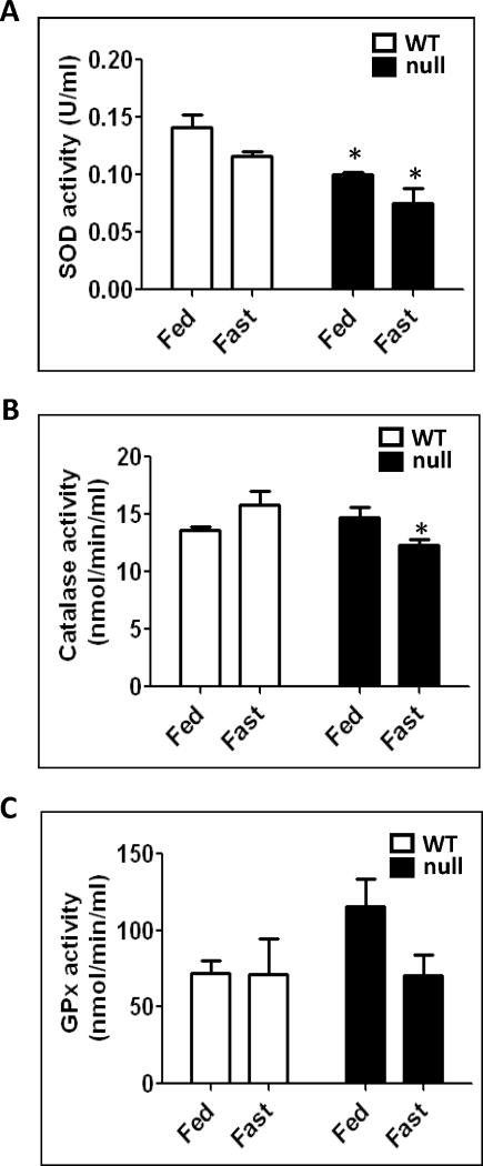
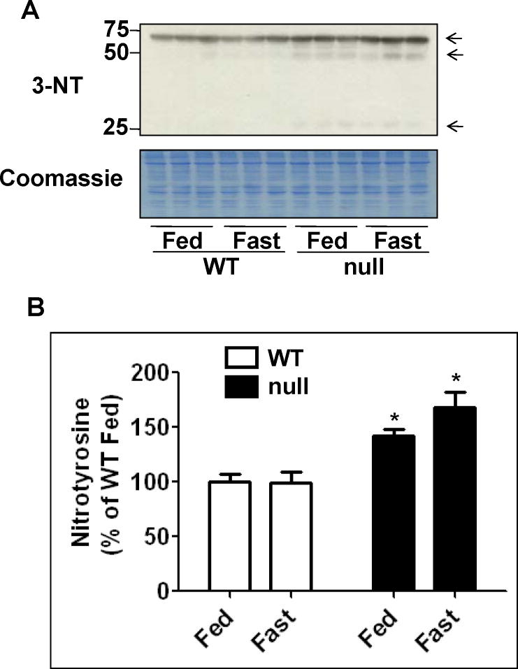
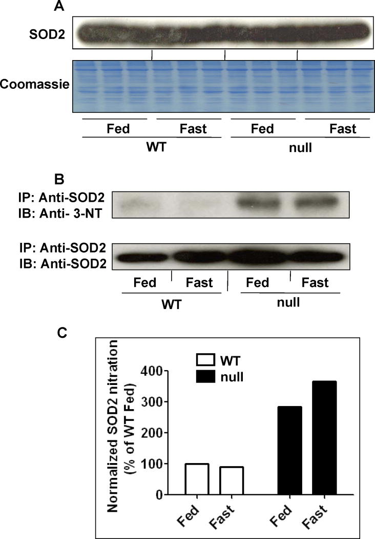
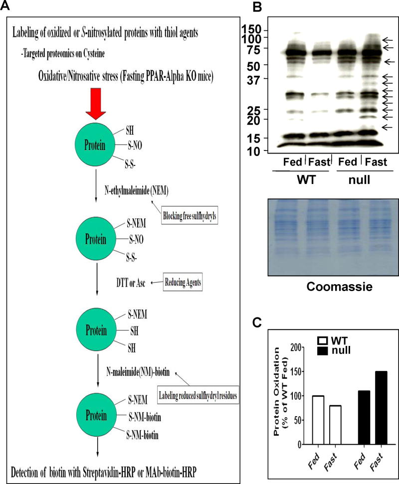
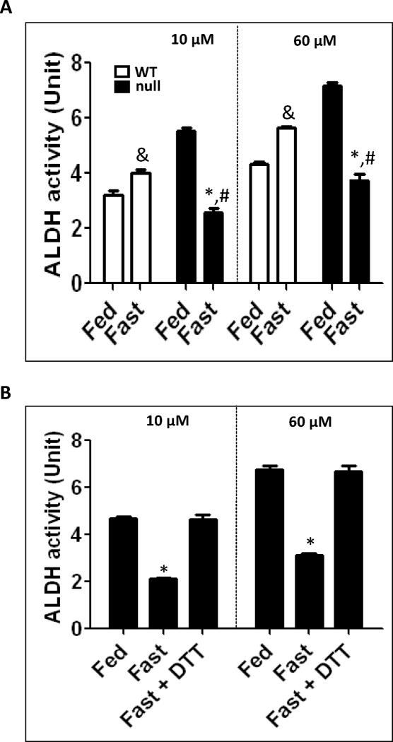
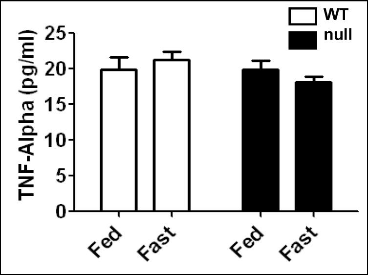
The peroxisome proliferator-activated receptor-alpha (PPARalpha) regulates lipid homeostasis, particularly in the liver. This study was aimed at elucidating the relationship between hepatosteatosis and oxidative stress during fasting. Fasted Ppara-null mice exhibited marked hepatosteatosis, which was associated with elevated levels of lipid peroxidation, nitric oxide synthase activity, and hydrogen peroxide accumulation. Total glutathione (GSH), mitochondrial GSH, and the activities of major antioxidant enzymes were also lower in the fasted Ppara-null mice. Consequently, the number and extent of nitrated proteins were markedly increased in the fasted Ppara-null mice, although high levels of protein nitration were still detected in the fed Ppara-null mice while many oxidatively modified proteins were only found in the fasted Ppara-null mice. However, the role of inflammation in increased oxidative stress in the fasted Ppara-null mice was minimal based on the similar levels of tumor necrosis factor-alpha change in all groups. These results with increased oxidative stress observed in the fasted Ppara-null mice compared with other groups demonstrate a role for PPAR alpha in fasting-mediated oxidative stress and that inhibition of PPAR alpha functions may increase the susceptibility to oxidative damage in the presence of another toxic agent.


Figures

Fig. 1
Fig. 1
Effect of fasting on intracellular lipids accumulation in wild-type and Ppara-null mice. Wild-type and Ppara-null mice were either fed or deprived of food for 36 h with free access to water. Removal of standard rodent chow was started at the beginning of the light cycle and mice were euthanized at the beginning of the dark cycle on the next day. Photomicrographs (200×) after H&E staining from the left hepatic lobes of indicated mouse livers are presented: (A) Wild-type fed standard chow, (B) Ppara-null fed standard chow, (C) Fasted wild-type, and (D) Fasted Ppara-null mice. Fasted wild-type mice (C) were normal as compared to Ppara-null-fasted mice (D) which showed pleomorphic foamy hepatocytes due to accumulation of micro- and macro-vesicular lipid droplets. Photomicrographs are representative of different treatment groups (n = 3 per group), and all mice exhibited a very similar pattern of response.
Fig. 2
Fig. 2
Effect of 36 h fasting on plasma transaminase activities. The activities of ALT and AST were measured in plasma samples from each animal in different groups by using a clinical chemistry analyzer system and presented. Data are expressed as mean ± S.E.M. of 3 mice per group.
Fig. 3
Fig. 3
Changes in body and liver weights of wild-type and Ppara-null mice following 36 h fasting. (A) Body weight of each mouse was measured before and after 36 h feeding or fasting with free access to water. The changes of weight are presented as percentage of weight reduction. (B) When mice were euthanized, the whole liver was excised and immediately weighed. Liver weights are presented as percentage of the body weight. Data are expressed as mean ± S.E.M. of 3 mice per group. *Significantly different from corresponding wild type group; #significantly different from fed Ppara-null mice.
Fig. 4
Fig. 4
Effect of 36 h fasting on CYP2E1 activity, MDA formation, NOS activity, H2O2 production, and total and mitochondrial GSH. Equal amounts of whole liver lysates or mitochondrial proteins from different groups were used to determine (A) CYP2E1 activity by measuring the rate of PNP oxidation to p-nitrocatechol, (B) hepatic malondialdehyde (MDA), (C) NOS activity, (D) H2O2 production rates, (E) total GSH and (F) mitochondrial GSH levels, as described in Materials and Methods. Data are expressed as mean ± S.E.M. of 3 mice per group. &Significantly different from fed wild-type; *significantly different from corresponding wild-type group; #significantly different from fed Ppara-null mice.
Fig. 5
Fig. 5
Changes in the anti-oxidant enzyme activities following 36 h fasting in wild-type and Ppara-null mice. Equal amounts of whole liver lysates were used to measure: (A) superoxide dismutase activity, (B) catalase activity, and (C) glutathione peroxidase activity according to the manufacturer’s protocols. Data are expressed as mean ± S.E.M. of 3 mice per group. *significantly different from corresponding wild-type group.
Fig. 6
Fig. 6
Levels of hepatic protein nitration in wild-type and Ppara-null mice subjected to fasting for 36 h. (A) Equal amounts of whole liver lysates (40 μg/well) from different groups were separated on 12% SDS–PAGE, transferred to nitro-cellulose membrane, and subjected to immunoblot analysis (IB) by using anti-3-NT antibody (upper panel). Coomassie-stained gel is presented to show equal protein loading. (B) Density of 3-NT band in each lane was calculated by quantitative densitometry, normalized to that of the corresponding Coomassie-stained protein bands, and is presented as a percentage of the 3-NT level detected in the wild-type fed group. Data are expressed as mean ± S.E.M of 3 mice per group. *significantly different from corresponding wild-type group.
Fig. 7
Fig. 7
Levels of mitochondrial superoxide-dismutase 2 (SOD2) and nitrated (SOD2) in wild-type and Ppara-null mice following 36 h of food deprivation. (A) Equal amounts of whole liver lysates (40 μg protein/well) from different groups were separated on 12% SDS–PAGE, transferred to nitro-cellulose membrane, and subjected to immunoblot analysis (IB) by using the specific anti-SOD2 antibody (upper panel). Coomassie blue-stained gel is presented to show equal protein loading (lower panel). (B) Whole liver lysates (1 mg/sample) were pooled from 3 mouse livers per group for the 4 different groups and were immunoprecipitated (IP) with the specific anti-SOD2 antibody, as described in the “Methods” section. Immunoprecipitated proteins from each group were then subjected to 12% SDS–PAGE, transferred to nitrocellulose membrane, and subjected to immunoblot analysis with the anti-3-NT (upper panel) or anti-SOD2 antibody (lower panel). (C) Density of 3-NT bands was normalized to that of the corresponding SOD2 band and is plotted as a percentage of the nitro-tyrosine level detected in the wild-type fed group.
Fig. 8
Fig. 8
Comparison of oxidized proteins in the four groups following 36 h fasting. (A) Schematic diagram of the method used to identify oxidatively-modified Cys residues. (B) Whole liver lysates were pooled from 3 mouse livers per group (10 mg/group) and labeled with N-EM. The N-EM-labeled proteins were then treated with 15 mM DTT for 30 min to reduce the oxidatively-modified Cys residues before they were incubated with biotin-N-maleimide. Equal amounts of biotin-labeled protein (10 μg/well) were separated on 12% SDS–PAGE, transferred to nitro-cellulose membrane, and subjected to immunoblot analysis with the anti-biotin antibody (upper panel). Coomassie blue-stained gel is presented to show equal protein loading (lower panel). (C) Densities of oxidized-protein bands were normalized to that of the corresponding Coomassie blue-stained bands and are plotted as a percentage of the oxidized-protein level in the wild-type fed group.
Fig. 9
Fig. 9
Inactivation of ALDH in fasted Ppara-null mice for 36 h. (A) Catalytic activities of mitochondrial ALDH2 (by using 10 μM propionaldehyde) and cytosolic ALDH1 (by using 60 μM propionaldehyde) in the indicated liver samples were determined. (B) Reversal of the suppressed mitochondrial ALDH2 and cytosolic ALDH1 from the fed- or fasted- Ppara-null mouse livers was determined in the absence and presence of 15 mM DTT. &Significantly different from fed wild-type; *significantly different from corresponding wild-type group (A) or from other groups (B); #significantly different from fed Ppara-null mice.
Fig. 10
Fig. 10
Effect of 36 h fasting on tumor necrosis factor-alpha (TNF-α) levels in wild-type and Ppara-null mice. Equal amounts of whole liver lysates were used to measure the contents of TNF-α by ELISA according to the manufacturer’s protocol and findings are presented.

References

    1. Kliewer SA, Forman BM, Blumberg B, Ong ES, Borgmeyer U, Mangelsdorf DJ, Umesono K, Evans RM. Differential expression and activation of a family of murine peroxisome proliferator-activated receptors. Proc Natl Acad Sci U S A. 1994;91:7355–7359. - PMC - PubMed
    1. Braissant O, Foufelle F, Scotto C, Dauca M, Wahli W. Differential expression of peroxisome proliferator-activated receptors (PPARs): tissue distribution of PPAR-alpha, -beta, and -gamma in the adult rat. Endocrinology. 1996;137:354–366. - PubMed
    1. Greene ME, Blumberg B, McBride OW, Yi HF, Kronquist K, Kwan K, Hsieh L, Greene G, Nimer SD. Isolation of the human peroxisome proliferator activated receptor gamma cDNA: expression in hematopoietic cells and chromosomal mapping. Gene Expr. 1995;4:281–299. - PMC - PubMed
    1. Desvergne B, Wahli W. Peroxisome proliferator-activated receptors: nuclear control of metabolism. Endocr Rev. 1999;20:649–688. - PubMed
    1. Mandard S, Muller M, Kersten S. Peroxisome proliferator-activated receptor alpha target genes. Cell Mol Life Sci. 2004;61:393–416. - PMC - PubMed

Publication types

MeSH terms

LinkOut - more resources