. 2009 Jul 14;19(13):1086-95.
doi: 10.1016/j.cub.2009.05.056. Epub 2009 Jun 18.

Compression regulates mitotic spindle length by a mechanochemical switch at the poles

Affiliations

Compression regulates mitotic spindle length by a mechanochemical switch at the poles

Sophie Dumont et al. Curr Biol. .

Abstract

Background: Although the molecules involved in mitosis are becoming better characterized, we still lack an understanding of the emergent mechanical properties of the mitotic spindle. For example, we cannot explain how spindle length is determined. To gain insight into how forces are generated and responded to in the spindle, we developed a method to apply controlled mechanical compression to metaphase mitotic spindles in living mammalian cells while monitoring microtubules and kinetochores by fluorescence microscopy.

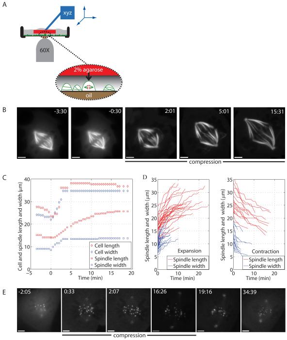
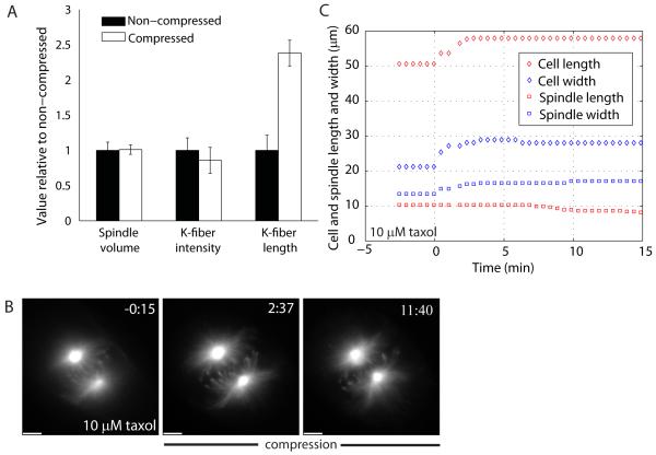
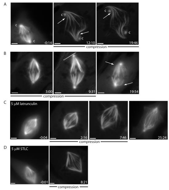
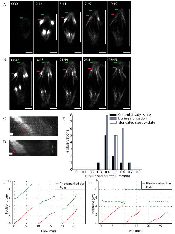
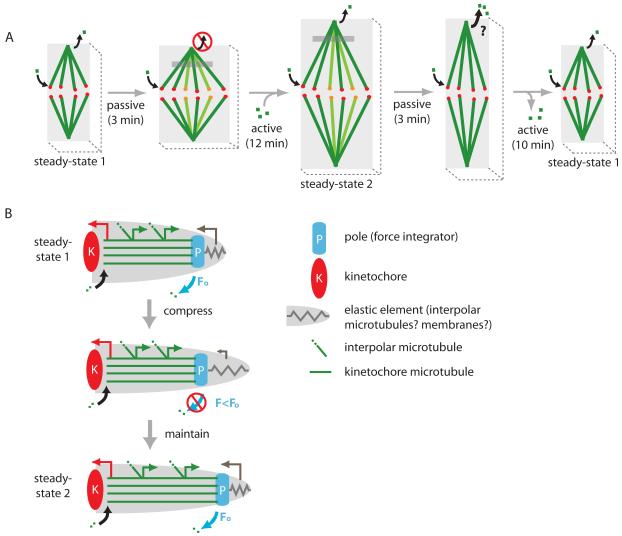
Results: Compression caused reversible spindle widening and lengthening to a new steady state. Widening was a passive mechanical response, and lengthening was an active mechanochemical process requiring microtubule polymerization but not kinesin-5 activity. Spindle morphology during lengthening and drug perturbations suggested that kinetochore fibers are pushed outward by pole-directed forces generated within the spindle. Lengthening of kinetochore fibers occurred by inhibition of microtubule depolymerization at poles, with no change in sliding velocity, interkinetochore stretching, or kinetochore dynamics.

Conclusions: We propose that spindle length is controlled by a mechanochemical switch at the poles that regulates the depolymerization rate of kinetochore fibers in response to compression and discuss models for how this switch is controlled. Poleward force appears to be exerted along kinetochore fibers by some mechanism other than kinesin-5 activity, and we speculate that it may arise from polymerization pressure from growing plus ends of interpolar microtubules whose minus ends are anchored in the fiber. These insights provide a framework for conceptualizing mechanical integration within the spindle.


Figures

Figure 1
Figure 1
A compressed mitotic spindle expands asynchronously and reversibly. (A) Method developed to mechanically perturb spindles in vivo. (B) Fluorescence imaging of a compressed spindle in a Ptk2 EGFP-α-tubulin cell. (C) Time courses of cell and spindle length and width changes upon compression for the cell in (B). (D) Time courses of spindle length and width changes during expansion (31 cells) and contraction (17 cells). For clarity, steady-state time points are not displayed. (E) Fluorescence imaging of a compressed spindle in a Ptk2 EYFP-cdc20 cell, with compression released at 16:30. (B-E), compression started at 0:00 (min:s) and scale bar corresponds to 5 μm.
Figure 2
Figure 2
Tubulin polymerization is required for spindle elongation. (A) Histogram comparing non-compressed and compressed spindle steady-states: spindle volume (n=7), k-fiber cross-sectional intensity (n=19) and length of k-fibers (cell in Fig. S1B), all normalized to the non-compressed value. (B) Fluorescence imaging of a Ptk2 EGFP-α-tubulin bipolar spindle being compressed in 10 μM taxol, with its (C) time course of cell and spindle length and width changes. Compression started at 0:00 (min:s) and scale bar corresponds to 5 μm.
Figure 3
Figure 3
Spindle elongation is driven by forces internal to the spindle that are kinesin-5-independent. (A) Example of extreme spindle compression where spindle poles disconnect from and grow passed centrosomes (marked ‘c’, with arrows marking k-fiber bends). (B) Example of a spindle elongating until it reaches the cell cortex, with cortex marked by a dashed line using the phase image (release at 10:00; arrows mark k-fiber bends post-release, suggesting that k-fibers impede shortening). Spindle being compressed in (C) 5 μM latrunculin (note bent interpolar microtubules at 7:46; release at 15:27) and in (D) 5 μM STLC. Ptk2 EGFP-α-tubulin cells, compression started at 0:00 (min:s) and scale bar corresponds to 5 μm.
Figure 4
Figure 4
Spindle compression reduces microtubule depolymerization at poles while leaving the tubulin sliding rate unchanged. Tubulin sliding (A) during and (B) after elongation in Ptk1 PA-GFP-α-tubulin spindles being compressed. Green bars mark the poles and red bars the leading edge of photomarked tubulin. Vertical white bar marks spindle length. Tubulin photomarking was performed at 2:25 (min:s) and 14:25. Compression started at 0:00 and scale bar corresponds to 5 μm. Kymographs show the motion of photomarked tubulin with respect to the poles (C) during (over 10 min) and (D) after (over 7.5 min) elongation; scale bar corresponds to 1 μm. (E) Histogram of tubulin sliding rates before (control), during and after spindle elongation. Respective positions of the photomark and pole in three example traces (F) during and (G) after elongation (time translation for clarity).
Figure 5
Figure 5
Compression regulates spindle size. (A) Sketch of the response of a Ptk2 spindle to compression. Curved black arrows depict tubulin polymerization at the kinetochore and depolymerization at the poles (poleward sliding). In panel 2, new k-fibers are in focus (different color). Gray region depicts a photomarked tubulin population. Depolymerization at poles is inhibited during elongation; we predict it to increase during contraction but this has not been shown. For simplicity, active and passive responses are depicted in series, while they actually occur in parallel. (B) Mechanical coupling model for spindle length regulation, where the length of K-MTs is determined by force-dependent effects on microtubule dynamics at poles. Coupling between tension/compression on poles and K-MT dynamics is provided either by force-dependent regulation of the activity of a depolymerase, or by a direct effect of force on depolymerization. In either case, we postulate that the depolymerization rate at poles responds to the sum [43] of all the forces exerted on K-MTs (F), that we divide into pulling on plus-ends by kinetochores (red arrow), an outwards sliding force that is generated along the length of the K-MTs (green arrows), and pushing on minus-ends by other parts of the spindle or cell (gray arrow). K-MTs grow at the sliding rate until the original F (Fo) returns, when depolymerization resumes and a new spindle length steady-state is reached.

Similar articles

Cited by

References

    1. Inoué S, Salmon ED. Force generation by microtubule assembly/disassembly in mitosis and related movements. Molecular Biology of the Cell. 1995;6:1619–1640. - PMC - PubMed
    1. Li X, Nicklas RB. Mitotic forces control a cell-cycle checkpoint. Nature. 1995;373:630–632. - PubMed
    1. Sharp DJ, Rogers GC, Scholey JM. Microtubule motors in mitosis. Nature. 2000;407:41–47. - PubMed
    1. Inoué S, Sato H. Cell motility by labile association of molecules. The nature of mitotic spindle fibers and their role in chromosome movement. Journal of General Physiology. 1967;50:259–292. - PMC - PubMed
    1. Pickett-Heaps JD, Forer A, Spurck T. Traction fibre: toward a “tensegral” model of the spindle. Cell Motility and the Cytoskeleton. 1997;37:1–6. - PubMed

Publication types

MeSH terms

LinkOut - more resources