. 2009 Jul 14;19(13):1075-85.
doi: 10.1016/j.cub.2009.05.057. Epub 2009 Jun 18.

The carboxyl terminus of Brca2 links the disassembly of Rad51 complexes to mitotic entry

Affiliations

The carboxyl terminus of Brca2 links the disassembly of Rad51 complexes to mitotic entry

Nabieh Ayoub et al. Curr Biol. .

Abstract

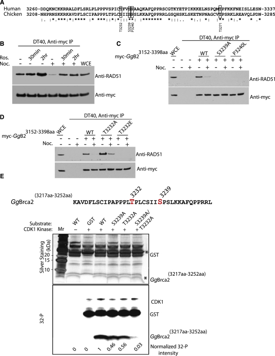
Background: The Rad51 recombinase assembles on DNA to execute homologous DNA recombination (HR). This process is essential to repair replication-associated genomic lesions before cells enter mitosis, but how it is started and stopped during the cell cycle remains poorly understood. Rad51 assembly is regulated by the breast cancer suppressor Brca2, via its evolutionarily conserved BRC repeats, and a distinct carboxy (C)-terminal motif whose biological function is uncertain. Using "hit-and-run" gene targeting to insert single-codon substitutions into the avian Brca2 locus, we report here a previously unrecognized role for the C-terminal motif.

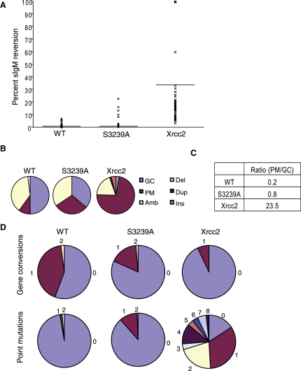
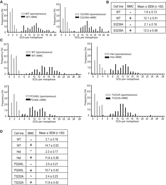
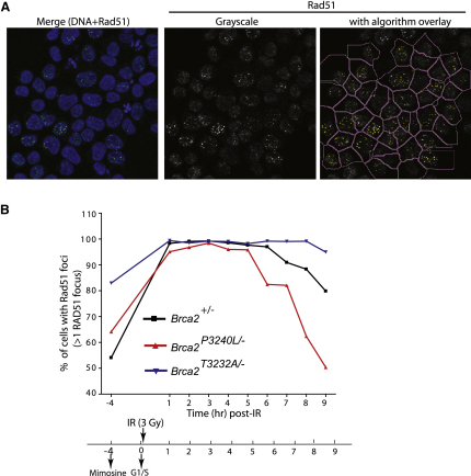
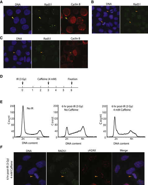
Results: We show that the avian C-terminal motif is functionally cognate with its human counterpart and identify point mutations that either abolish or enhance Rad51 binding. When these mutations are introduced into Brca2, we find that they affect neither the assembly of Rad51 into nuclear foci on damaged DNA nor DNA repair by HR. Instead, foci disassemble more rapidly in a point mutant that fails to bind Rad51, associated with faster mitotic entry. Conversely, the slower disassembly of foci in a point mutant that constitutively binds Rad51 correlates with delayed mitosis. Indeed, Rad51 foci do not persist in mitotic cells even after G2 checkpoint suppression, suggesting that their disassembly is a prerequisite for chromosome segregation.

Conclusions: We conclude that Rad51 binding by the C-terminal Brca2 motif is dispensable for the execution of HR but instead links the disassembly of Rad51 complexes to mitotic entry. This mechanism may ensure that HR terminates before chromosome segregation. Our findings assign a biological function for the C-terminal Brca2 motif in a mechanism that coordinates DNA repair with the cell cycle.


Figures

Figure 1
Figure 1
The C-Terminal Motif of GgBrca2 Is Functionally Cognate with Its Human Counterpart in CDK-Regulated Rad51 Binding (A) Protein sequence alignment showing the conserved cyclin-dependent kinase (CDK)-phosphorylated residues flanking Ser3239 of GgBrca2 and Ser3291 of HsBRCA2. Gallus gallus residues used in this study are boxed, and residue numbers are indicated below. Asterisks indicate identical residues, double dots indicate conserved substitutions, and single dots indicate residues that are semiconserved. (B) Western blot analysis of GgRad51 and the myc epitope following immunoprecipitation of the myc-tagged construct encoding residues 3152–3398 of GgBrca2 (GgB23152 aa–3398 aa) transfected into DT40 cells. Roscovitine treatment for 30–120 min in asynchronous cells had little effect on GgRad51 binding (lanes 2 and 3). However, roscovitine effectively reversed the reduction in binding induced by nocodazole (compare lanes 5 and 6 with lane 4). (C and D) Western blot analysis of GgRad51 and the myc epitope following immunoprecipitation comparing the wild-type (WT) with S3239A/E or P3240L (C) or T3232A (D) variants of GgB23152 aa–3398 aa transfected into DT40 cells. The S3239A/E and P3240L mutations in the conserved Ser-Pro consensus for CDK phosphorylation abrogated binding to GgRad51 in asynchronous and nocodazole-arrested mitotic cell extracts, whereas the T3232A mutation caused enhanced binding to GgRad51 under the same conditions. (E) Thr3232 and Ser3239 can be phosphorylated in vitro by CDK1. A wild-type GgBrca2 peptide fused with GST (WT, sequence at top) or mutant forms in which Thr3232 (T3232A), Ser3239 (S3239A), or both (T3232A/S3239A) were substituted with Ala were subjected to an in vitro kinase assay in the presence of [γ-32P]ATP. Reaction products were cleaved with thrombin to separate the Brca2 peptides from GST. Silver staining (middle panel) measures the loading of the peptides in each reaction (). The bottom panel shows that CDK1 catalyzes the transfer of 32P radiolabel from [γ-32P]ATP to the T3232A or S3239A peptides, but not to the double-mutant T3232A/S3239A peptide. Numbers at bottom indicate relative phosphorylation normalized to the amount of peptide present in each sample.
Figure 2
Figure 2
Immunoglobulin Gene Diversification in the Mutant Cell Line Brca2S3239A/S3239A Confirms the Integrity of Homologous DNA Recombination (A) Luria-Delbrück fluctuation analysis shows no significant difference in sIgM+ reversion to sIgM− status between the WT and S3239A cell lines. Xrcc2-deficient cells show a significant increase in median fluctuation relative to WT and S3239A (median percentage reversion indicated by horizontal line: WT 0.67%, S3239A 0.57%, Xrcc2 17.6%). (B) Pie charts showing that the source of mutation in VL1 is predominantly gene conversion in WT and Brca2S3239A/S3239A cells but predominantly point mutation in Xrcc2-deficient cells (n = 149, 148, and 54, respectively). (C) Table showing the ratio of point mutation to gene conversion suggests that commitment to homologous DNA recombination (HR) (gene conversion) is intact in Brca2S3239A/S3239A cells but has shifted to nontemplated error-prone repair pathways (point mutation) in Xrcc2-deficient cells. (D) Pie charts showing frequencies of unique gene conversion and point mutation events in WT and Brca2S3239A/S3239A cells indicate a preference for HR over the point mutation used extensively in Xrcc2-deficient cells.
Figure 3
Figure 3
No Defect in Spontaneous or Mitomycin C-Induced Sister Chromatid Exchanges in the Brca2S3239A/S3239A, Brca2+/−, Brca2P3240L/−, and Brca2T3232A/− Cell Lines (A) Frequency distribution histograms of spontaneous (gray) and mitomycin C (MMC)-induced (black) sister chromatid exchanges (SCEs) in WT and Brca2S3239A/S3239A cell lines show significant induction in both (two-tailed unpaired t test, n = 50). (B) The mean ± SEM of each frequency distribution in (A) is indicated. Contrary to prediction, no significant diminution of basal or MMC-induced SCEs in Brca2S3239A/S3239A cells was observed. Fold induction from SCE levels in unchallenged cells is 6.7 in WT compared to 6.3 in S3239A mutant. (C) Frequency distribution histograms of spontaneous (gray) and MMC-induced (black) SCEs in WT, Brca2+/−, Brca2P3240L/−, and Brca2T3232A/− cell lines show a significant induction of SCE by MMC in all cell lines (two-tailed unpaired t test, n = 50). Both Brca2T3232A/− and Brca2P3240L/− cells exhibit basal and MMC-induced SCE comparable to their parental heterozygous Brca2+/− cells. (D) Table enumerating SCEs (mean ± SEM) in unchallenged or MMC-treated cells. Heterozygous cells and mutants derived from them have basal levels of SCEs similar to WT cells, but modestly less induction after MMC treatment. Fold inductions are: WT, 7.0; Het, 5.1; P3240L, 4.4; T3232A, 5.0.
Figure 4
Figure 4
Normal Formation but Altered Dissolution of Damage-Induced Rad51 Foci in Brca2P3240L/− and Brca2T3232A/− Cells (A) Representation of a spot-counting algorithm on the Cellomics ArrayScan VTI. Left panel: a merged image of DNA (blue) and Rad51 (green). Middle and right panels: grayscale image of Rad51 exported to the Cellomics ArrayScan platform with (middle) and without (right) the algorithm overlay (details in Experimental Procedures). (B) Kinetics of Rad51 focus formation and dissolution in mimosine-synchronized Brca2+/−, Brca2P3240L/−, and Brca2T3232A/− cells after exposure to 3 Gy ionizing radiation (IR). Five hundred cells were analyzed on the Cellomics ArrayScan for each data point, and the percentage of cells positive for Rad51 foci was plotted as a function of time (see Experimental Procedures for details). Nearly 100% of cells formed Rad51 foci after IR in all three cell lines by 3 hr after damage. However, decreased binding to Rad51 in the Brca2P3240L/− cell line correlated with a faster reduction in focus-positive cells compared to the heterozygous control. Conversely, the Brca2T3232A/− cell line showed a slower reduction in foci dissolution.
Figure 5
Figure 5
Rad51 Foci Are Absent from Mitotic Cells (A) Immunofluorescence analysis of asynchronous WT DT40 cells costained for Rad51 (green) and cyclin B (red). A typical prophase cell (yellow arrowhead) shows intense nuclear (but not cytosolic) staining of cyclin B and is negative for Rad51 foci. DNA is stained with DAPI (blue). (B and C) Immunofluorescence staining of asynchronous human U2OS cells for RAD51 (green) and/or cyclin B (red). RAD51 foci are absent from U2OS cells marked by condensed DAPI-stained chromosomes that have entered mitosis (orange arrowheads). (D) Schematic of the experimental timeline. (E) Cell-cycle profiles of asynchronous, irradiated, and irradiated plus caffeine-treated U2OS cells. DNA content measured by propidium iodide staining and flow cytometry is plotted on the horizontal axis against relative cell number. 2n and 4n peaks represent the G1 and G2/M phases, respectively. (F) Representative immunofluorescence micrographs confirming the absence of RAD51 foci (green) in a mitotic cell (arrowhead) in which γH2AX staining (red) persists after exposure to IR and caffeine. DNA was stained with DAPI (blue). No Rad51 foci were observed in any of 50 mitotic nuclei identified by DAPI staining, whereas γH2AX staining persisted in 38 (76%) of them.
Figure 6
Figure 6
Coordinate Regulation of Mitotic Entry with Rad51 Binding by the C-Terminal Brca2 Motif (A) Brca2+/−, Brca2P3240L/−, and Brca2T3232A/− cells were synchronized with mimosine at the G1/S boundary and released into S phase. Samples collected at the indicated time points were stained with anti-MPM2 and analyzed by flow cytometry. The percentage of MPM2-positive cells is plotted on the vertical axis against time after release from G1/S. Each data point represents a mean ± SEM percentage from three samples. MPM2 staining marks cells entering mitosis, which usually increases 4 hr after release from G1/S in control Brca2+/− parental cells (Figure S7B). In the Brca2P3240L/− cell line, this increase is hastened, whereas conversely, in the Brca2T3232A/− cell line, it is delayed. (B) A hypothetical model for the biological role of the C-terminal Brca2 motif in coordinating DNA repair by HR with mitotic entry. During the G2 phase, phosphorylation of the C-terminal Brca2 motif by CDKs regulates the disassembly of DNA-bound Rad51 complexes (1), a process monitored by a mechanism that is independent of the G2 checkpoint for DNA damage (2). Completed disassembly allows entry into the M phase.

Similar articles

Cited by

References

    1. Branzei D., Foiani M. Interplay of replication checkpoints and repair proteins at stalled replication forks. DNA Repair (Amst.) 2007;6:994–1003. - PubMed
    1. Cox M.M. The nonmutagenic repair of broken replication forks via recombination. Mutat. Res. 2002;510:107–120. - PubMed
    1. Kogoma T. Stable DNA replication: Interplay between DNA replication, homologous recombination, and transcription. Microbiol. Mol. Biol. Rev. 1997;61:212–238. - PMC - PubMed
    1. Li X., Heyer W.D. Homologous recombination in DNA repair and DNA damage tolerance. Cell Res. 2008;18:99–113. - PMC - PubMed
    1. Haber J.E. DNA recombination: The replication connection. Trends Biochem. Sci. 1999;24:271–275. - PubMed

Publication types

MeSH terms